
Figure 4.
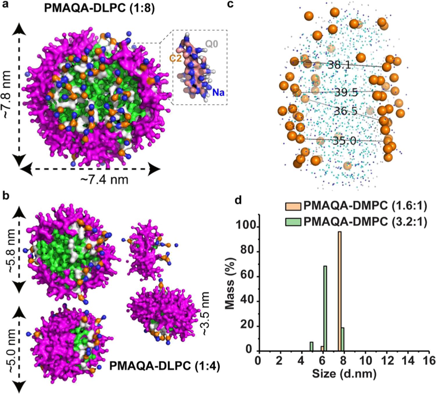
MD snapshots showing the formation of the DLPC nanodisc by ~4.8 kDa PMAQA at a variable polymer to lipid ratio. Spontaneous formation of nanodiscs by ~4.8 kDa PMAQA at 1:8 (a) and 1:4 (b) polymer to DLPC ratio at 10 μs MD simulation. The polymers are shown in violet and DLPC atoms in different colors as indicated in Figure 2. Water molecules and ions are not shown for transparency. (c) Distance between the lipid head groups (in Å) of PMAQA nanodiscs shown in (a) is measured using PyMOL. (d) DLS showing the size distribution of PMAQA–DLPC nanodiscs at the indicated polymer to lipid (w/w) ratio.