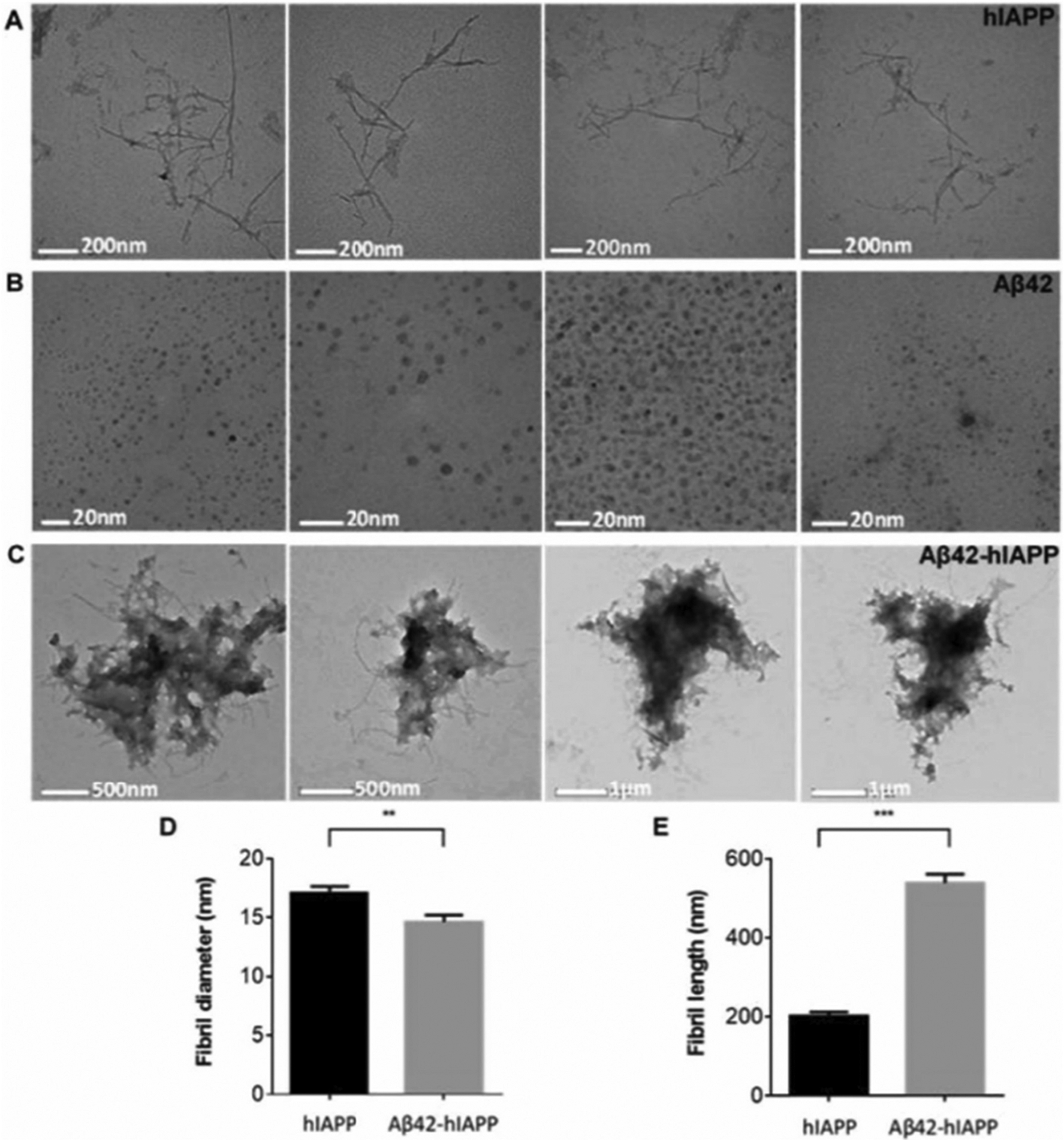
Fig. 3.

Electron micrographs of hIAPP, Aβ42 and Aβ42-hIAPP samples showing (A) fibril-like hIAPP (17 ± 0.5 nm in width, 202 ± 9.9 nm in length, n = 77) and spherical (B) Aβ42 oligomers (6 ± 0.2 nm in diameter, n = 207). (C) Aβ42-hIAPP formed large amorphous aggregates. Analysis of fibril (D) length and (E) diameter demonstrated that Aβ42-hIAPP (14 ± 0.6 nm in diameter, 539 ± 22.7 nm in length, n = 100) was significantly different from hIAPP (mean ± SEM, **p < 0.005, ***p < 0.001). Aβ42-hIAPP mixtures are large amorphous structures that are distinctly different from either spherical Aβ42 oligomers or fibril-like hIAPP. Scale bar (A) 200 nm, (B) 20 nm, (C) 500 nm/μm. The figure was adapted with permission from Reference [74].
