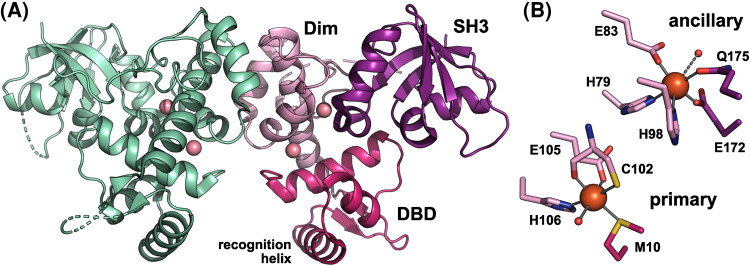
Figure 1. The structure of IdeR in the metallated state.
(A) The overall structure of the Co2+-activated SeIdeR dimer (PDB 7B1V) [11], with one subunit colored by domain and the Co2+ ions shown as pink spheres (DBD, DNA-binding domain; Dim, dimerization domain; SH3, SH3-like domain). (B) Detailed view of the metal-binding sites, showing the Fe2+- (and DNA-)bound state of SeIdeR (PDB 7B20) [11], using the same coloring scheme as in A (orange spheres, Fe2+ ions; small red spheres, water molecules). Cys102 co-ordinates the metal ion with its sulfur as well as its backbone carbonyl oxygen atoms. The water molecule in the ancillary site only weakly co-ordinates the metal ion (3.6 Å distance), as indicated by the dashed line. This figure was prepared with PyMOL (Schrödinger, LLC).

