Fig. 1.

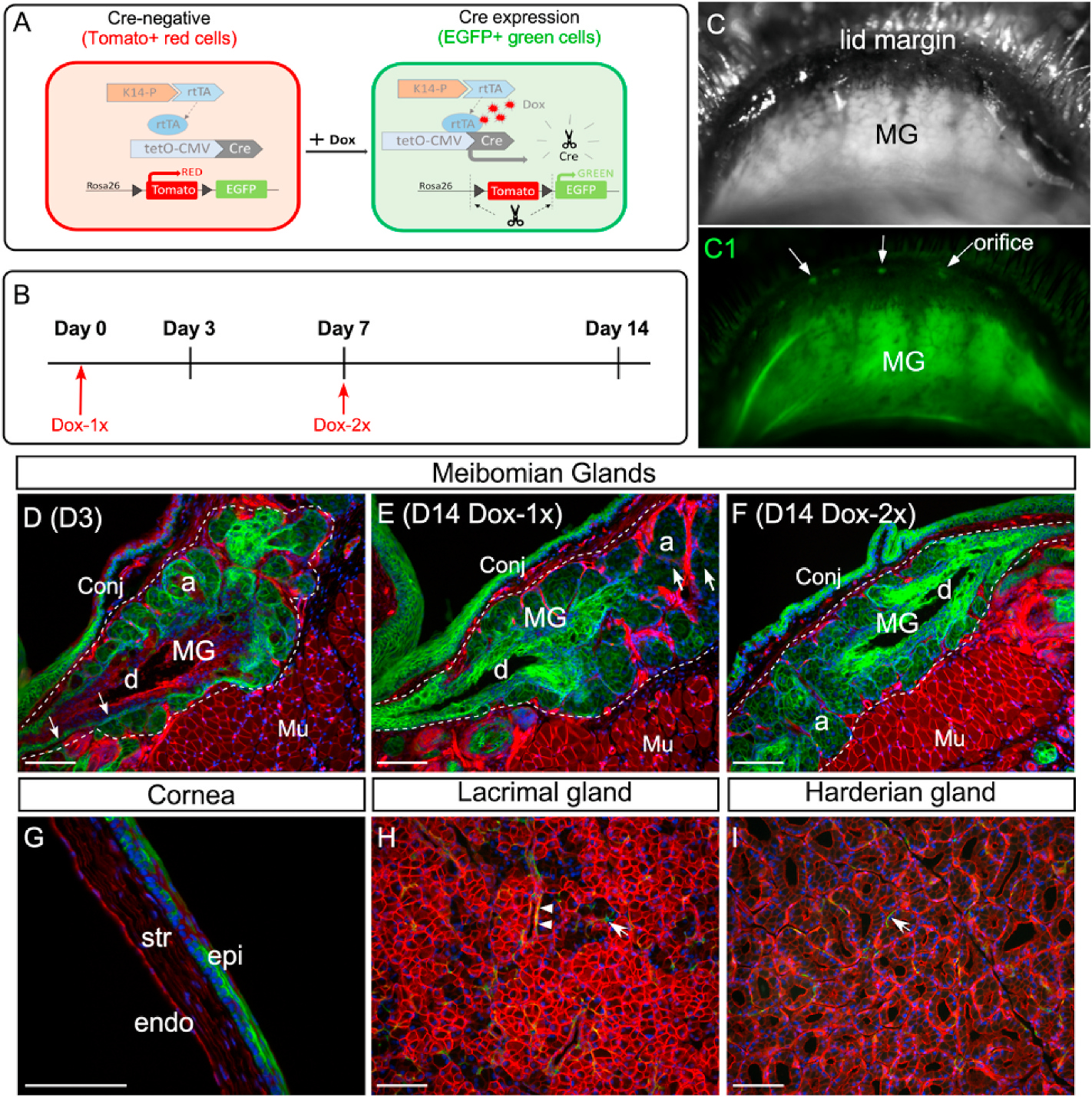
Dox-induced EGFP expression in the ocular surface tissues and glands of the reporter mice. (A) Illustration of the dual fluorescence reporter system in the triple transgenic mice (K14rtTA;tetOCre; RosamTmG). In the absence of Dox, all cells express Tomato (red) fluorescence. Upon Dox induction, Cre recombinase is activated by reverse tetracycline-controlled transactivator (rtTA) driven by the K14 promoter, resulting in the expression of EGFP (green fluorescence) in the K14-positive cells. (B) Experimental scheme. (C and C1) Live imaging of MGs in a reporter mouse after 14-day chase from the first Dox injection. The MGs were shown under regular (C) and fluorescent (C1) illumination. Arrows point to the MG orifice. (D–E) Cryosections of upper eyelids from Dox-injected reporter mice stained with nuclear dye DAPI (blue). The MG area was outlined by dotted line. On day 3 (D3) after the first injection, EGFP fluorescence was readily seen in the acini (a) and was weakly visible in the basal epithelium of the central duct (d) (indicated by arrows in D). After 14-day chase, EGFP fluorescence was diminished in some acini (arrows in E) as a result of the holocrine secretion process in MGs. In the central duct, EGFP expression was expanded from the basal to the suprabasal layers during the chase from D3 (D) to D14 (E). EGFP was also expressed in the conjunctival epithelium (conj) as early as D3. (F) When two sequential doses of Dox was given on D1 and D7 (Dox-2x, as illustrated in B), EGFP fluorescence was present in most of the acini on D14. (G) Mosaic EGFP expression in the corneal epithelium on D14 in the Dox-2x reporter mice. (H–I) Weak EGFP expression in the lacrimal gland (H) and Harderian gland (I) after 14-day chase. A few EGFP-positive myoepithelial cells (arrows in H and I) and ductal cells (arrowheads in H) were identified in these glands. Abbreviations: a, acini; d, duct; MG, meibomian gland; conj, conjunctiva; epi: corneal epithelium; str: stroma; endo: corneal endothelium; Mu: Muscle. Scale bar in each figure represents 100 μm.
