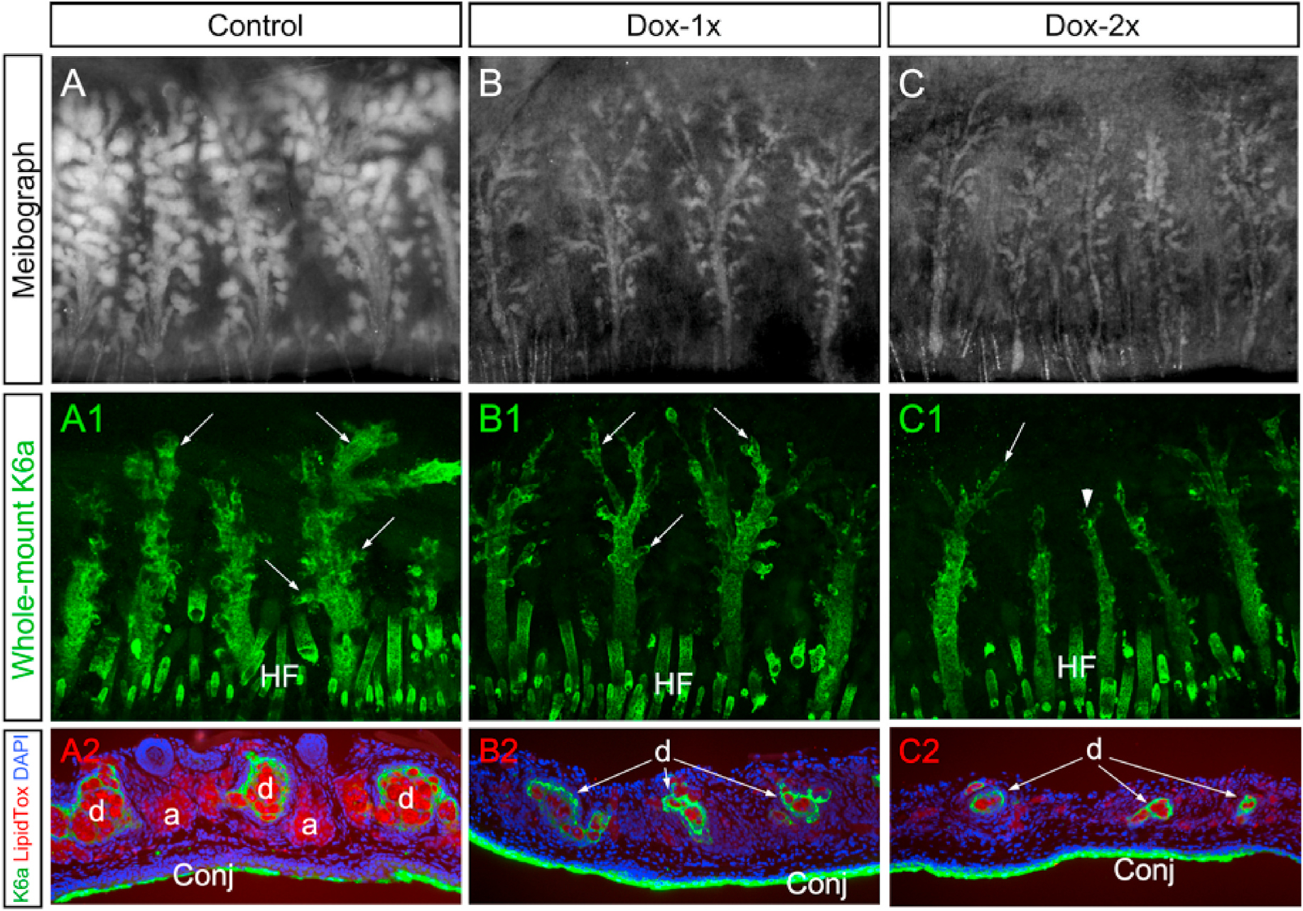
Fig. 3.

MG acinar and ductal atrophy shown respectively by meibography and whole-mount K6a immunofluorescence staining of tarsal plates. (A–C) Meibographs showed normal (A) and atrophic acini in Dox-1x (B) and Dox-2x (C) Fgfr2CKO mice on D14 after the first Dox-injection. (A1-C1) Corresponding to the meibographs shown in A-C, whole-mount K6a immunofluorescence (green) of tarsal plates exhibited progressive loss and atrophy of the ductal tissues in the Dox-injected Fgfr2CKO mice (B1 and C1) when compared with the control (A1). The ductal branches (arrows) were either attenuated (B1 and C1) or completely lost in the distal end of the MGs (arrowhead in C1) in the Dox-injected Fgfr2CKO mice. (A2-C2) The whole-mount K6a-stained tarsal plates were processed for cryosections and co-stained with LipidTOX (red) to further demonstrate both acinar and ductal atrophy in the Dox-injected Fgfr2CKO mice, which occurred more severely in the Dox-2x than in the Dox-1x mice. Abbreviation: HF, hair follicle.
