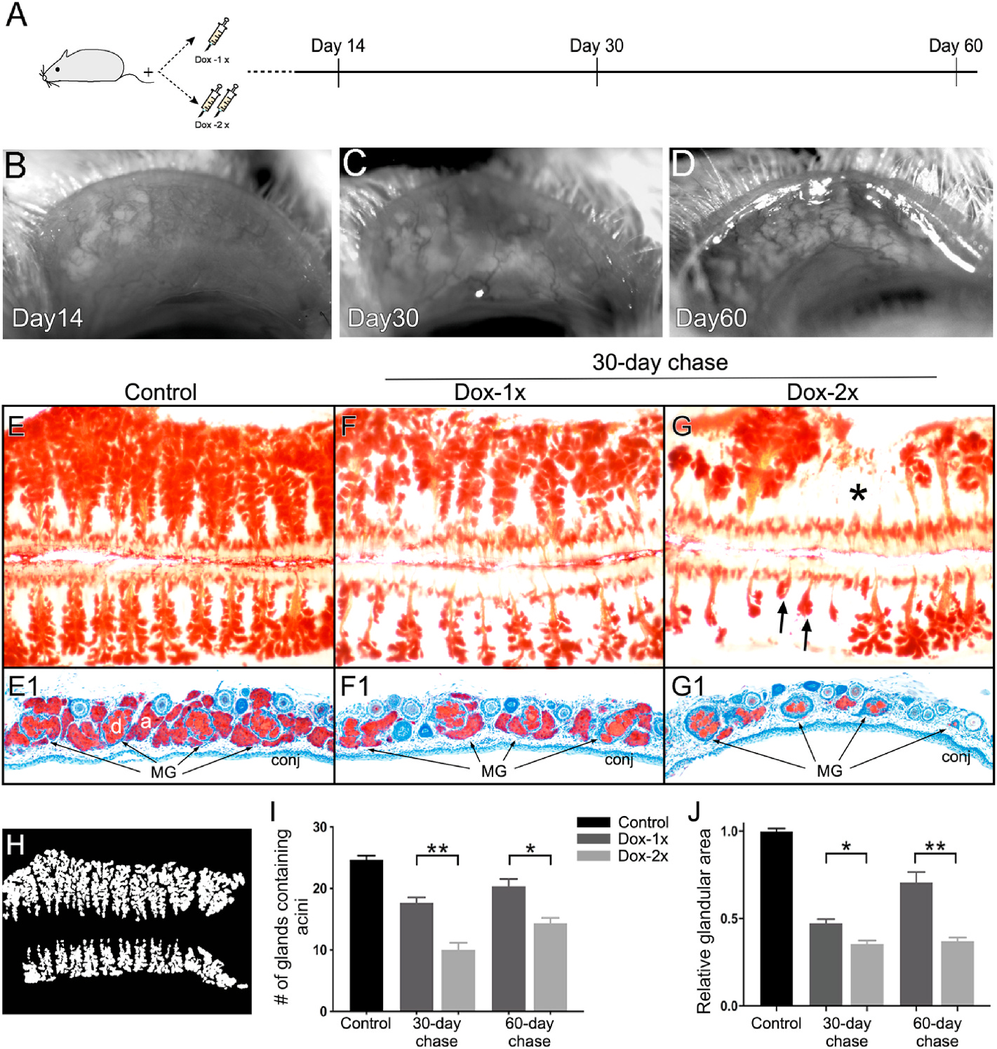
Fig. 4.

Spontaneous glandular recovery after Dox-induced atrophy in Fgfr2CKO mice. (A) Experimental scheme to examine MG recovery from atrophy. (B–D) In vivo imaging showing severe MG atrophy on D14 (B) followed by progressive acinar regeneration on D30 (C) and D60 (D) in a Dox-1x Fgfr2CKO mouse. (E–G) ORO whole mount staining of paired eyelids in the control (E), Dox-1x (F) and Dox-2x (G) Fgfr2CKO mice after 30-day chase. Gland dropout (asterisk in G) and shortening (arrows in G) were noted in the Dox-2x mouse. (E1-G1) Cryosections of the corresponding ORO-stained tarsal plates counter-stained with hematoxylin to confirm the different degree of glandular recovery from atrophy in the Dox-1x and Dox-2x mice. (H–J) Quantitative analysis of MG acinar tissue recovery. The ORO-stained meibographs were converted into the binary images for quantitative analysis (H). The number of MGs containing lipid-producing acini (I) and the total recovered glandular area (J) were calculated. Reduced acinar recovery was associated with more severe ductal damage in the Dox-2x Fgfr2CKO mice. Data was shown as mean ± SEM. n = 3 mice per group, *p < 0.05, and **p < 0.01.
