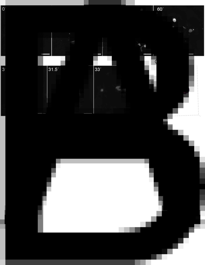
Fig. 4.
Besnoitia besnoiti bradyzoites induced vital NETosis. Live cell 3D holotomographic microscopy (Nanolive®) analysis under controlled temperature and atmosphere conditions was performed for 1 h of interactions registering images every 30 s (A). At 31 min of incubation a tossing vital NETosis is observed without compromising the overall structure of PMN (B). Digital staining and 3D holotomographic reconstruction of tossed vital NETosis (C). (A) Scale bar = 20 μm, (B) Scale bar = 10 μm.

