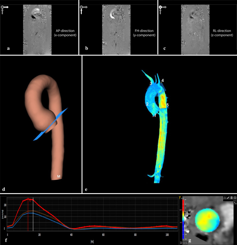
Fig. 1.
General workflow of analyzing aortic 4D flow MRI data. a–c depict the phase images of the phase contrast data with velocity encoding in three directions (i.e. anterior–posterior (AP; X-component), feet-head (FH; Y-component), and left–right (LR, Z-component). After the pre-processing steps, including phase offset and anti-aliasing correction, luminal segmentation is performed on the combined phase contrast images. This results in a 3D model as seen in image d. Image e represents the streamlines of blood flow within the aortic volume. Manual placement of 2D planes along the centerline results in a quantified flow graph (f). Each line represents a 2D plane and a cross-sectional image of through plane velocity profile (g). Red depicts high velocity; blue depicts low and retrograde velocity

