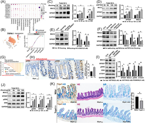
FIGURE 6.

The role of TRPA1 in intestinal goblet cells. (A) Dot plot showing the expression of mechanosensitive ion channels in intestinal epithelial cells. (B) t‐Stochastic neighbor embedding (tSNE) plot showing the expression of TRPA1 in epithelial cells; the expression of TRPA1 in goblet cells of the functional and defunctioned intestines (***p < .001; ns = not significant, Wilcoxon rank‐sum test). (C and D) Mechanical stimulation and TRPA1‐selective agonist (ASP7663, 5 μM) promoted the expression of SPDEF and MUC2 (n = 3). All cells were cultured for 24 h and treated with a rocking board (Rocking) for mechanical stimulation or ASP7663 for 6, 12 and 24 h, respectively. (E) Treatment with TRPA1‐selective inhibitor (HC‐030031, 5 μM) blocked the expression of SPDEF and MUC2 induced by mechanical stimulation (n = 3). (F) Blocking the expression of TRPA1 inhibited the expression of SPDEF and MUC2 induced by mechanical stimulation (n = 3). (G) Gene set enrichment analysis showed enriched RAS signal in goblet cells of the functional intestine. (H) Immunohistochemistry staining of pERK in functional and defunctioned intestines (*p < .05, n = 5, scale bars = 50 μm, paired Wilcoxon rank‐sum test). (I) Treatment with ASP7663 (5 μM) promoted the expression of pERK and treatment with ERK selective inhibitor (PD98059, 10 μM) inhibited the expression of SPDEF and MUC2 induced by ASP7663; the expression of ATOH1 was not affected (n = 3). (J) Mechanical stimulation promoted the expression of SPDEF, MUC2 and pERK (n = 3). All cells were cultured for 24 h, and cells were mechanically stimulated using a Rocking for 6, 12 and 24 h, respectively. (K) Immunohistochemistry staining for Trpa1, HE staining and alcian blue (AB) staining of intestine in wild‐type and Trpa1−/− mice (*p < .05, n = 5, scale bars = 50 μm, Wilcoxon rank‐sum test). (C–F) and (I–J); *p < .05, **p < .01, ns = not significant, unpaired t‐test. All values are presented as mean ± SEM of each group. Con, control.
