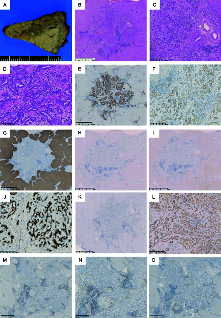
Fig. 2.
Macroscopic and histological findings of the liver segment 3 (S3) tumor. The S3 tumor was a light-green 11×6×7 mm nodule (A). Histopathological examination revealed neoplastic small bile ductules (cholangiocytes) arranged in a staghorn-like configuration similar to bile ductular proliferation (hematoxylin and eosin staining) (B: magnification ×20; C: magnification ×100). The S3 tumor appeared as cells with round to oval nuclei that proliferated in an anastomosing pattern of small glands without mucus production, mimicking ductular reactions with a background of abundant fibrous stroma (D: magnification ×200). This tumor was immunohistologically positive for cytokeratin (CK) 7 (E: magnification ×20) and CK19 (F: magnification ×200) and negative for hepatocyte markers, such as hepatocyte paraffin 1 (Hep Par 1) (G: magnification ×20), glypican 3 (H: magnification ×20), and CD10 (I: magnification ×20). Epithelial membrane antigen was positivity at the glandular lumen (J: magnification ×200). Neither p53-immunopositive cells (K: magnification ×20) nor nuclear expression of β-catenin was observed (L: magnification ×100). Infiltrating CD8-positive T-lymphocyte were observed (M: magnification ×20) (N: magnification ×40) and CD4-positive T-lymphocytes were not observed (O: magnification ×20).

