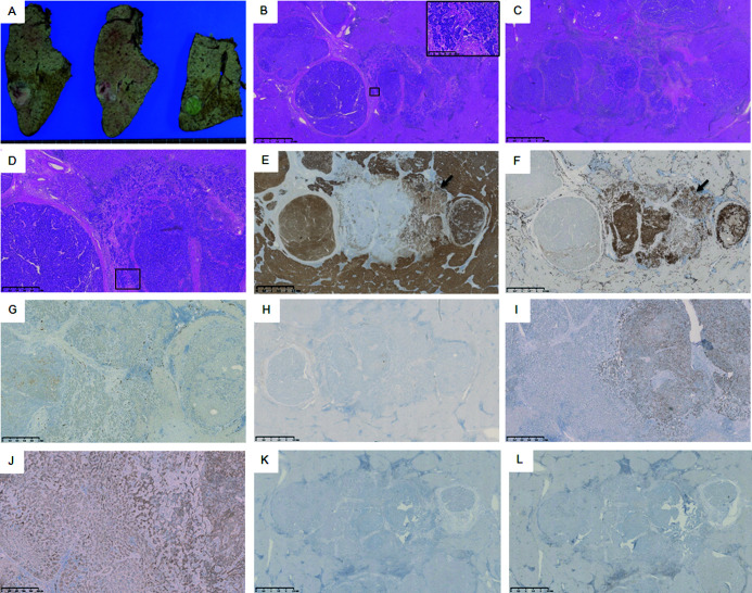
Fig. 3.
Macroscopic and histological findings of the liver segment 6 (S6) tumor. The S6 tumor appeared as an off-white and green 38 x 23 x 33 mm. nodule (A). The tumor consisted of both moderately differentiated hepatocellular carcinoma and moderately differentiated cholangiocarcinoma (hematoxylin and eosin staining B: magnification ×20; C: magnification ×20 of consecutive sections; D: magnification ×50). No cancerous emboli traces were found in the vein and lymph vessel of both tumors. The hepatic tissue adjacent to the tumors was cirrhotic. Hepatocellular carcinoma (HCC) with a thick trabecular appearance (lateral) and cholangiocarcinoma (CCA) with glandular structures were embedded in the desmoplastic stroma (central) and intimately interdigitated at the transitional region (B, C, and D). These CCA cells showed mucus production (box in B; magnification ×200). Immunohistochemistry revealed that the HCC component was positive for hepatocyte markers, such as hepatocyte paraffin 1 (Hep Par 1) (E: magnification ×20). The CCA component was positive for cytokeratin (CK) 7 (F: magnification ×20), partially positive for CK19 (G: magnification ×50), and negative for Hep Par1 (E: magnification ×20) and epithelial membrane antigen (H: magnification ×20). p53-immunopositive cells were observed in only the CCA component (I: magnification ×50). Nuclear expression of β-catenin was not observed (J: magnification ×100). Neither CD8-positive T-lymphocyte (K: magnification ×20) CD4-positive T-lymphocytes were not observed (L: magnification ×20).

