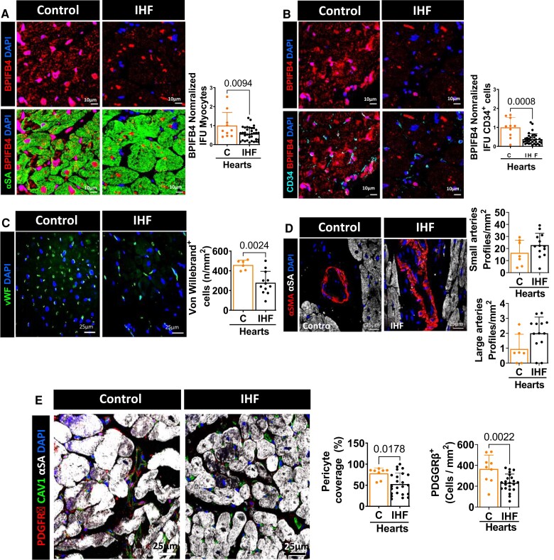
Figure 1.
Immunohistochemical characterization of human hearts. (A–B) Expression of BPIFB4 in cardiomyocytes (A) and endothelial cells (B) from controls and IHF hearts. (C–E) Microvascular alterations in IHF hearts. Capillary density is decreased in IHF compared with control hearts (C), whereas the reduction in arteriole density did not reach a statistical significance (D). PC density and coverage are lower in hearts explanted from elderly patients with IHF (E). PCs stained with PDGFRβ (red), endothelial with vWF or CAV1 (green) or CD34 (light blue) and cardiomyocytes with α-sarcomeric actin (αSA, green or white). Nuclei are identified by DAPI (blue) and BPIFB4 expression labelled in red. n = 8–9 C hearts and 23-22 IHF hearts. Data were analyzed using the Mann–Whitney U test (panels B, D, and E, pericyte coverage IHF vs. C) or unpaired Student’s t-test (all the other panels).

