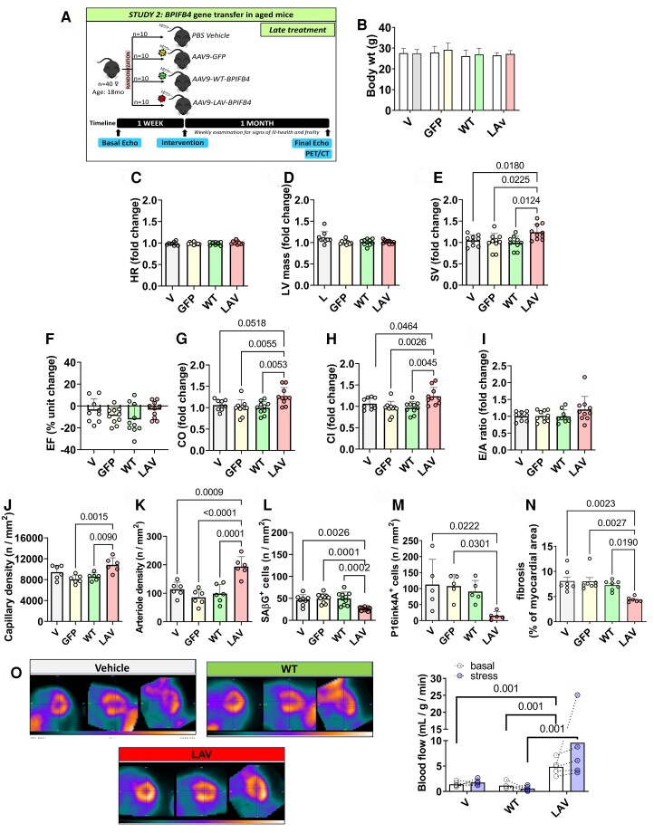
Figure 7.
Late LAV-BPIFB4 gene therapy improves cardiac function in elderly mice. (A) Cardiac function was assessed in female mice at baseline (18 months old) and four weeks post-treatment (19 months old). n = 10 female mice per group. (B) Body weight. (C–I) Fold changes in functional parameters from basal measurements except for Ejection Fraction, which is expressed as absolute unit change. Heart rate (HR) (C), left ventricular mass (D), stroke volume (SV) (E), ejection fraction (EF) (F), cardiac output (CO) (G), cardiac index (CI) (H), and E/A ratio (I). (J–K) Graphs show capillaries (J) and arteries (K) density. n = 6 mice per group. (L–M) Graphs showing data of the density of β-galactosidase (L) and p16ink4A (M) positive cells. n = 5 to 10 mice per group. (N) Cardiac fibrosis was assessed after staining with Azan Mallory in female mice at 1-month post-treatment (19 months old). n = 6–7 mice per group. (O) Representative images of PET imaging were performed in subgroups of the early and late intervention studies. Representative images. The bar graph shows the data from basal and Dobutamine stress tests. n = 5 mice per group. Values are presented as mean, standard deviation, and individual values. Data were analyzed using ANOVA followed by Tukey's multiple comparisons test, except for panels D and G where the Kruskal–Wallis test was applied followed by Dunn's multiple comparisons test.

