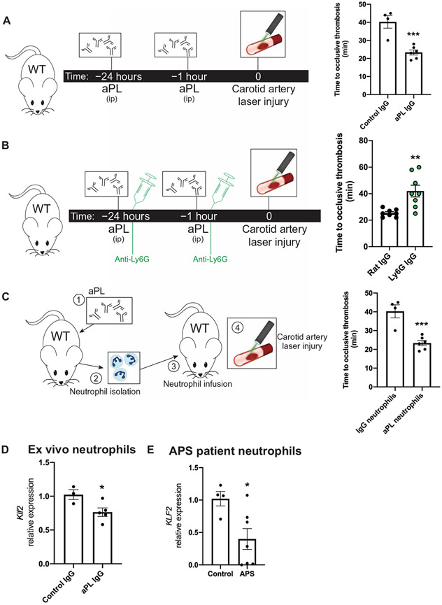
Fig. 1. Activated neutrophils drive arterial and venous thrombosis.
(A) In the murine antiphospholipid antibody (aPL)–injected model, mice were injected intraperitoneally (ip) with either aPL isolated from patients with APS (n = 6 mice) or IgG isolated from healthy controls (n = 4 mice) at 24 hours and 1 hour before a carotid artery laser-induced injury model of thrombosis. Time to occlusive thrombus development was measured. (B) Neutrophil depletion in aPL-treated mice using anti-Ly6G antibody (n = 8 mice) or control IgG (n = 8 mice) was followed by carotid artery injury to measure dependency on neutrophils in arterial thrombus formation. Time to occlusive thrombosis development was measured. All mice received aPLs. (C) Neutrophils were harvested from IgG- or aPL-treated wild-type (WT) mice and transferred into untreated WT mice (n = 4 IgG-treated neutrophil recipients; n = 6 aPL-treated neutrophil recipients) before carotid artery injury. (D) Klf2 expression was measured in neutrophils harvested from IgG-injected (n = 3) or aPL-injected (n = 5) WT mice. (E) KLF2 expression was measured in neutrophils harvested from humans with APS (n = 7) or healthy controls (n = 4). *P < 0.05, **P < 0.01, and ***P < 0.001 by unpaired, two-tailed Student’s t test.

