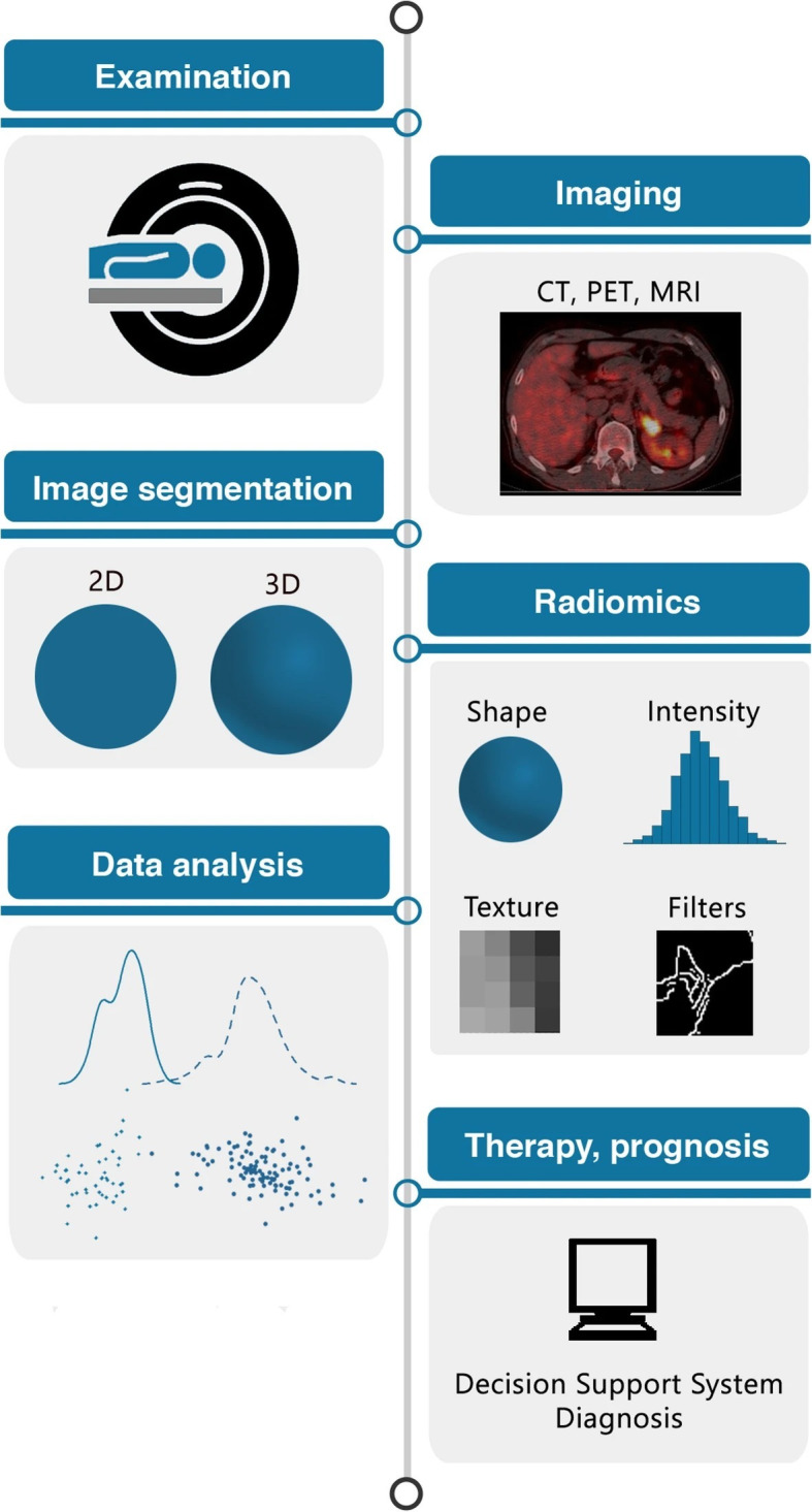
Figure 1.

The radiomics process and sequential steps, including image acquisition, segmentation of region of interest, extraction of radiomic features, and predictive modeling and subsequent validation.10 2D, 2-dimensional; 3D, 3-dimensional; CT, computed tomography; MRI, magnetic resonance imaging; PET, positron emission tomography. Source: Reprinted from van Timmeren JE, Cester D, Tanadini-Lang S, Alkadhi H, Baessler B. Radiomics in medical imaging—”how-to” guide and critical reflection. Insights Imaging. 2020;11:91. https://doi.org/10.1186/s13244-020-00887-2.10
