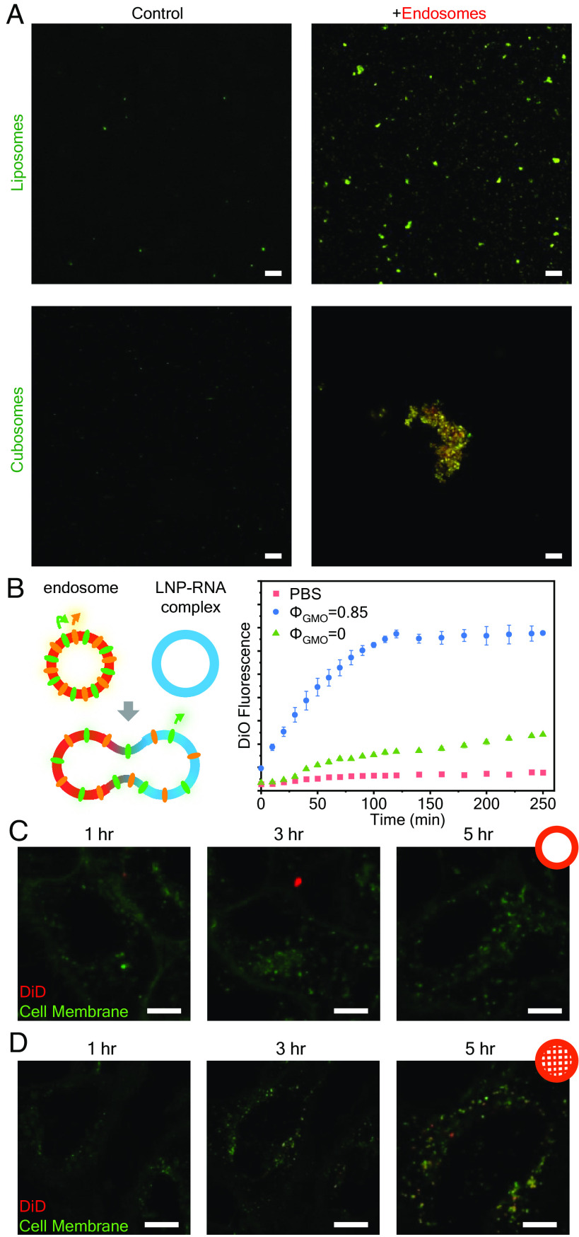
Fig. 4.
Cubic and hexagonal LNPs fuse more readily with endosomes compared to lamellar LNPs. (A) Visualization of LNP–endosome interaction with CLSM after 6 h incubation. (Scale bar, 20 μm.) (B) FRET assay to evaluate membrane fusion between endosomes and LNP–siRNA complexes of different structures. The fusion extent is indicated by DiO fluorescence recovery (n = 3, data presented as mean ± SD). (C and D) Live cell imaging of HeLa cells treated with lipoplexes (C) and cuboplexes (D) labeled with 1% self-quenching dye DiD. Recovery of DiD signal (red) implies LNP–endosomal fusion. (Scale bar, 10 μm.)

