Fig. 1.
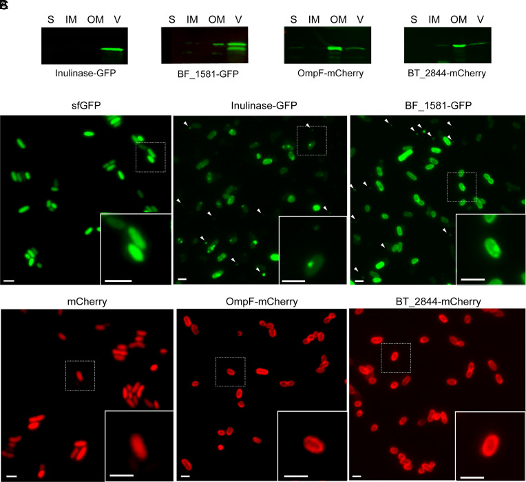
OMV and OM chimeric markers show a differential distribution in Bt. (A) Western blot of 10 μg of protein from soluble fraction (S), inner membrane (IM), outer membrane (OM), and OMV (V) fractions of Bt expressing Inulinase-GFP, BF_1581-GFP, OmpF-mCherry, or BT_2844-mCherry. Anti-His and anti-mCherry antibodies were employed to identify GFP and mCherry chimeric markers, respectively. (B) Representative widefield fluorescence microscopy images of OMV chimeric markers Inulinase-GFP and BF_1581-GFP. White arrows indicate the presence of OMVs. (C) Representative widefield fluorescence microscopy images of OM chimeric markers OmpF-mCherry and BT_2844-mCherry. Cytosolic expression of sfGFP and mCherry are used as reference (Left). (Scale bar: 2 μm.)

