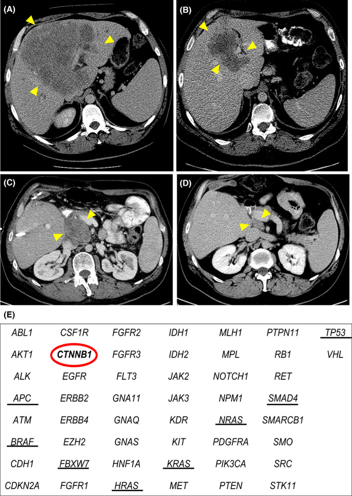
FIGURE 4.

Disease course in a patient who presented with colorectal liver metastases (CLM) centrally located across both hepatic lobes and the caudate lobe after previous resection of primary colon cancer. Yellow arrowheads indicate the tumor. (A) Computed tomography (CT) image at the time of the initial visit showing a large tumor centrally located extending to the hilar plate and caudate lobe. (B) CT image after preoperative chemotherapy showing partial response with persistent invasion of the hilar plate and caudate lobe. (C) CT image 7 mo after extended left hepatectomy with common bile duct and caudate lobe resection showing recurrence in a retro‐portal lymph node. (D) CT image after chemotherapy showing partial response. (E) Results of gene panel analysis of 50 genes. The red circle indicates CTNNB1 alteration of the tumor, and black underlines indicate driver genes associated with oncologic outcome after resection of CLM. Please note the absence of driver gene alteration in keeping with the good prognosis observed in this patient.
