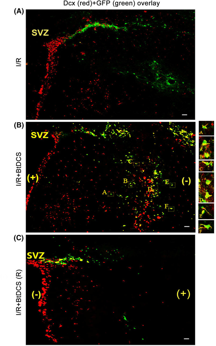
FIGURE 4.

Colabeling of Dcx with GFP‐labeled cells in the ipsilateral striatum of MCAO rat after BtDCS treatment. (A–B) Representative images show that BtDCS exposure of 150 μA for 30 min per day for 2 days after MCAO for 12 days, increases the colocalization of Dcx with GFP in ipsilateral striatum at 14 days after ischemia–reperfusion injury. Scale bar = 100 μm. (C) BtDCS exposure of 150 μA for 30 min per day for 2 days after MCAO for 12 days, with the anodal electrode placed on the ischemic hemisphere, decreases the colocalization of GFP with Dcx in the ischemic striatum at 14 days after ischemia–reperfusion injury. Scale bar = 100 μm.
