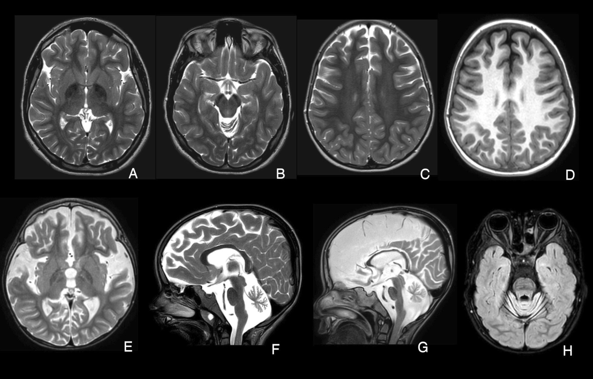
Fig. 2.

MRI-based imaging findings in patients. A-B) Patient with PLA2G6 due to c.2370 T > G and c.1516G > A variants; Axial T2-weighted images indicate hypointensity of the Globus pallidus (a) and substantia nigra (b) denoting iron deposition at the age of 11 years. c, d Patient with PLA2G6 due to c.668C > T variant; Axial T2 & T1-weighted images show white matter hypomyelination at the age of 4 years. e, f Patients with PLA2G6 due to c.668C > T variant and c.668C > T variant; Axial and sagittal T2-weighted images show cerebral atrophy and cerebellar atrophy at the age of 4 years. g Patient with PLA2G6 due to c.668C > T variant; Thinning of corpus callosum and calval hypertrophy at the age of 4 years. h Patient with PLA2G6 due to c.668C > T variant; The initial stage of optic atrophy on axial FLAIR sequence at the age of 4 years
