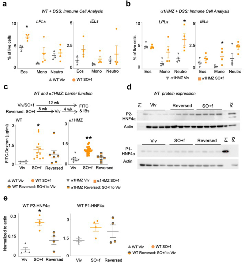
Figure 4.

A diet high in LA increases immune dysfunction and decreases barrier function in WT and α1HMZ mice.
Notes: Immune cell analysis (Eos, eosinophils; Mono, monocytes; Neutro, neutrophils) in DSS-treated WT (a) or α1HMZ (b) mice (see Supplementary Figure S2D and S3D for data from untreated mice); * vs WT or α1 HMZ Viv P < .05, T-test was performed between Viv and SO+f for each cell type, N = 3–4 per group. (c) Epithelial barrier permeability in WT and α1HMZ mice fed either Viv or SO+f diets for 12 weeks or SO+f for 8 weeks, followed by 4 weeks of Viv (reversed) (one outlier removed from WT reversed group). * vs Viv, ** vs Viv and Reversed. P < .05, one-way ANOVA, Sidak’s post hoc comparison. N = 6–22 per group. (d) HNF4α immunoblots of whole cell extracts (WCE, 30 µg) from distal colon of WT mice fed Viv chow or SO+f diet for 12 weeks or SO+f for 8 weeks followed by Viv chow for 4 weeks (reversed). Each lane contains WCE from a different mouse. P1 control-nuclear extract from HCT116 cells expressing P1-HNF4α; P2 control-nuclear extract from α7HMZ mouse (see Supplementary Figure S4 for entire blots for WT mice and P1 blot for α1HMZ mice). (e) Quantification of the P1-and P2-HNF4α immunoblot signals (shown in Figure 4d) normalized to total protein, as determined by actin staining of the same blot. * P < .05, one-way ANOVA, Tukey’s post hoc comparison. N = 3–4 per group.
