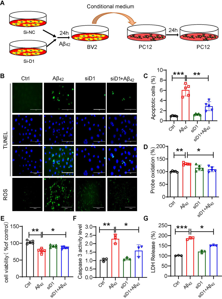
Figure 7.
Dectin-1 interference prevents Aβ42-induced neuronal damage. (A) BV2 cells pretreated with siD1 for 24 h are stimulated with/without 20 µM Aβ42 for another 24 h, and the conditioned culture supernatant is collected. (B) PC12 cells are plated into four groups: CTL, Aβ42, siD1, and siD1+Aβ42; BV2 serum is added to stimulate the PC12 cells for 24 h; and cell viability is measured by MTT assay. (C) LDH assay is used to test cell membrane damage. (D) TUNEL staining and Ros staining are used to test the apoptosis and intracellular ROS level [scale bar = 100 μm]. (E) Quantification of apoptotic cell in TUNEL staining in C. (F) Caspase 3 activity is detected using caspase 3 assay. (G) Quantification of ROS staining in D.

