
Fig. 1.
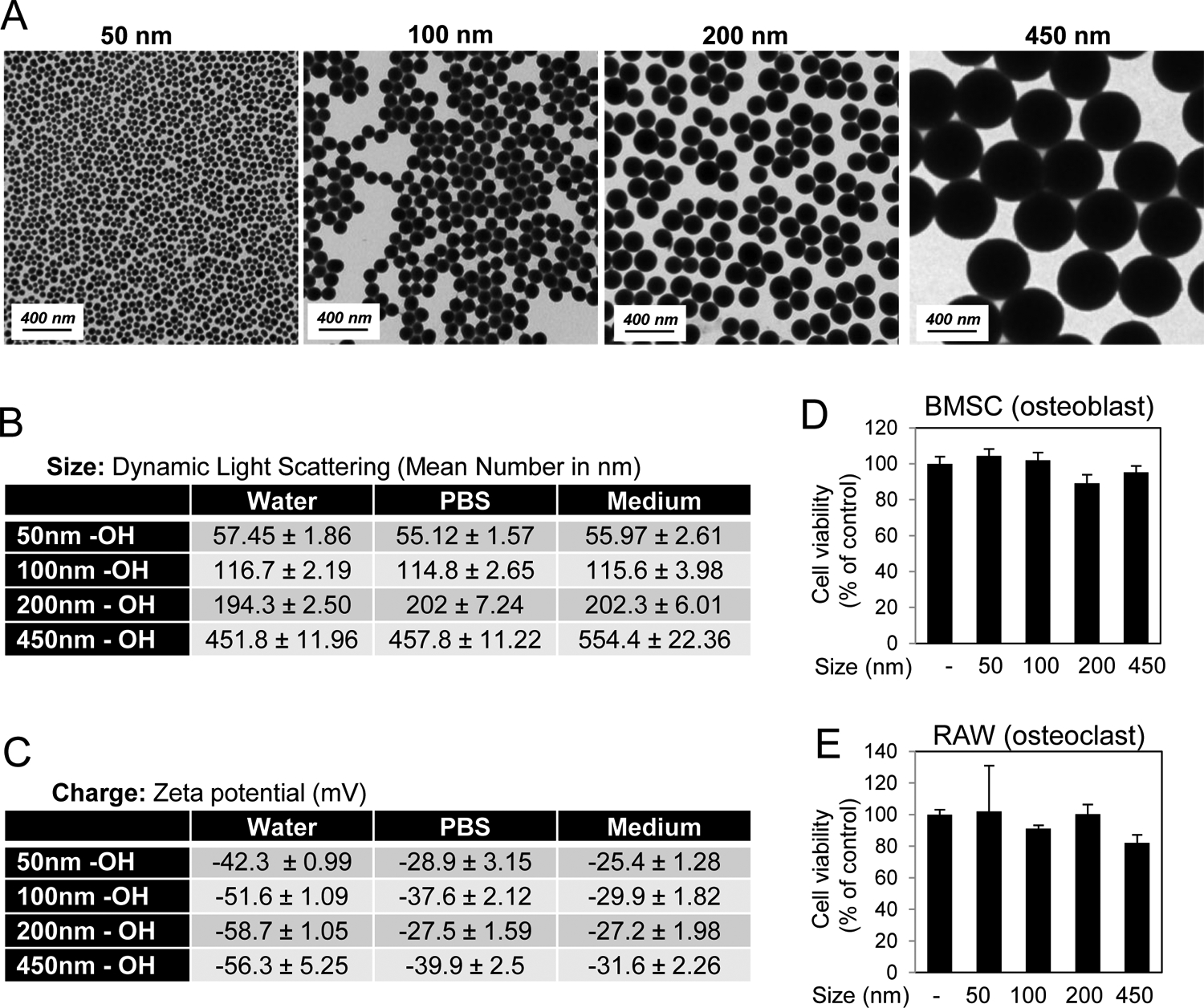
Characterization of varying sized silica nanoparticles. A) Shape was assessed by Transmission Electron Microscopy. B) Size of nanoparticles was characterized by Dynamic Light Scattering in water, phosphate buffered saline (PBS), and medium (Avg. of 3 readings). C) Zeta Potential of nanoparticles was measured in water, DPBS, and cell culture medium (Avg. of 3 readings). Cell viability of D) BMSCs in response to 72 hr treatment with 10 μg/ml nanoparticles and E) RAW264.7 cells in response to 72 hr treatment with 25 μg/ml nanoparticles as indicated (N=3–6). Avg.±Stdev.