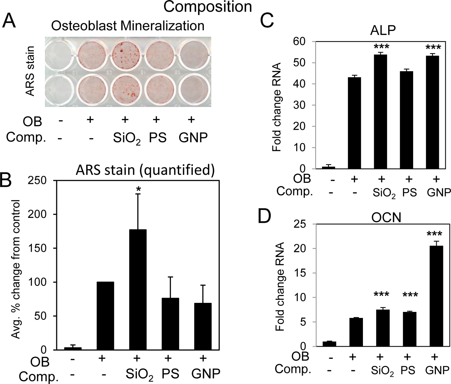
Fig. 8. Nanoparticle composition and enhancement of osteoblast differentiation.

BMSCs were cultured in growth medium or medium supplemented with ascorbic acid and beta glycerophosphate (OB) and treated with 10 μg/ml of 50 nm nanoparticles composed of SiO2:silica nanoparticles, GNP:gold nanoparticles and PS:polystyrene. Cells were stained after 11–14 days for A) mineralization with Alizarin Red S staining and B) the average of four independent experiments quantified by image J. Data are expressed and percent change from the positive control (+OB) ±Stdev. Gene expression of C) alkaline phosphatase (ALP) and D) osteocalcin (OCN) are expressed as fold change (±Stdev) relative to untreated. Statistical analysis compares nanoparticle treated relative to positive control (+OB) by Student’s t test. *P< 0.05, **P< 0.01, **P< 0.005.
