Figure 2.
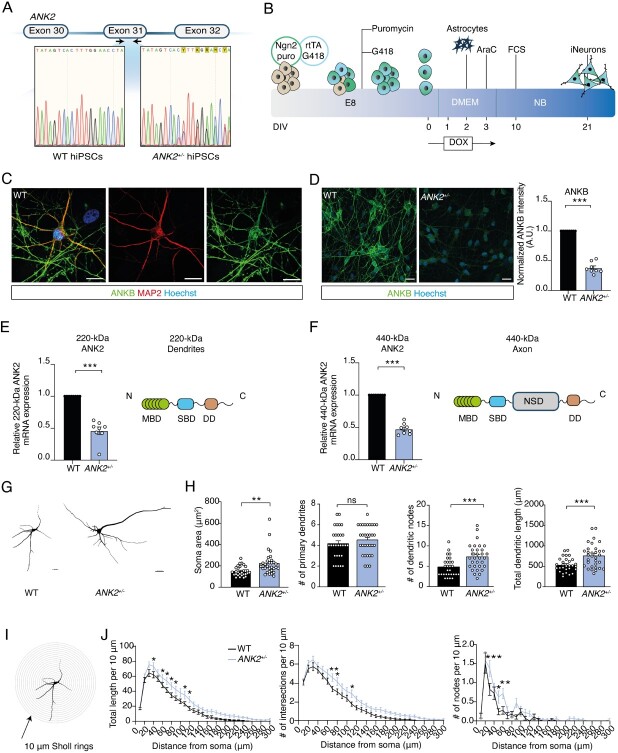
Heterozygous loss of 220-kDa and 440-kDa isoforms of ANK2 leads to reduced ANKB expression in iNeurons and increases somatodendritic structures. (A) Schematic diagram of CRISPR/Cas9 genetic editing in exon 31 and the DNA sequences before and after targeted gene editing. One nucleotide insertion causes a frameshift variant in WT hiPSCs. (B) Schematic presentation of the iNeuron differentiation protocol. (C) Representative images of a WT iNeuron immunostained for MAP2 (red) and ANKB (green) (scale bar 10 μm). (D) Representative images of ANK2+/− iNeurons immunostained for ANKB (green) (scale bar 5 μm), and quantification of ANKB fluorescent intensity, n = 8 for WT; n = 4 for ANK2+/− clone 1 and n = 4 for ANK2+/− for clone 2. ***P < 0.001 by two-tailed unpaired t-test. (E) Schematic presentation of protein domains expressed in 220-kDa ANKB, and qPCR analysis of 220-kDa ANK2 isoform performed in WT and ANK2+/− iNeurons at DIV 21. Values of ANK2+/− are normalized to WT (n = 8). ***P < 0.001 by two-tailed unpaired t-test. (F) Schematic presentation of protein domains expressed in 440-kDa ANKB, and qPCR analysis of 440-kDa ANK2 performed in WT and ANK2+/− iNeurons at DIV 21. Values of ANK2+/− are normalized to WT (n = 8). ***P < 0.001 by two-tailed unpaired t-test. (G) Representative somatodendritic reconstructions of WT and ANK2+/− iNeurons (scale bar 20 μm). (H) Quantification of the soma size, the number of primary dendrites, the number of dendritic nodes and the total dendritic length, n = 30 for WT; n = 30 for ANK2+/−. **P < 0.01 and ***P < 0.001 by two-tailed unpaired t-test. (I) Representative image of a WT iNeuron in sequential 10 μm rings placed from the center soma outwards for Sholl analysis. (J) Quantification per 10 μm Sholl section of the total dendritic length per ring, the number of dendritic intersections per ring and the number of dendritic nodes per ring, n = 30 for WT; n = 30 for ANK2+/−. *P < 0.05, **P < 0.001, ***P < 0.001 by two-way ANOVA with post hoc Bonferroni correction. MBD = membrane-binding domain; SBD = spectrin-binding domain; RD = regulatory domain.

