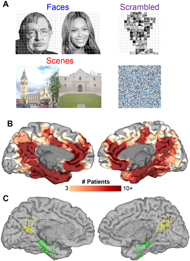
Figure 1. Task and Patients.

(A) Representative stimuli for each stimulus category. (B) Spatial coverage represented as a heat map on a standardized N27 brain surface showing extensive and consistent coverage of the medial parietal cortex and medial temporal lobe (n = 50 patients that performed both face and scene identification correctly). (C) Individual electrode locations (9,016 total electrodes) highlighting electrodes in the two ROIs defined using a parcellation derived from the Human Connectome Project [79]. Responsive electrodes are shown in MPC (yellow, n = 142) and MTL (green, n = 216). Electrodes within these ROIs not responsive to either faces or scenes are in maroon and electrodes outside these two ROIs are in white
