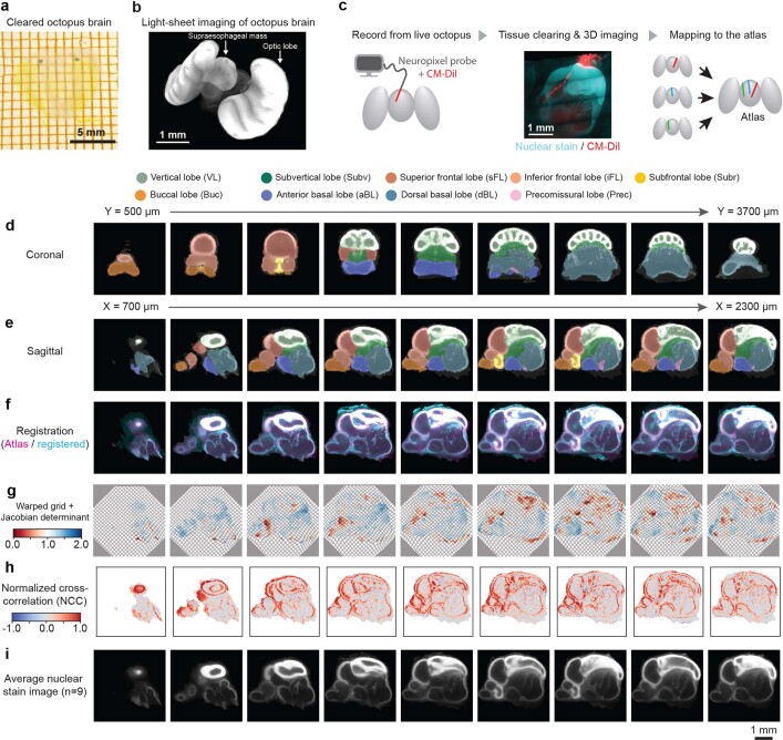
Extended Data Fig. 3. Octopus brain atlas and Neuropixels mapping.
a) Adult O. laqueus brain, cleared with CUBIC (Methods). b) 3D rendering of the cleared octopus brain imaged with a light sheet microscope. c) Neuropixels mapping workflow. Neuropixels probe was coated with CM-DiI to leave fluorescent labelling of penetration track in the brain. The brain was cleared and imaged using a light sheet microscope with dual channels (nuclear staining and CM-DiI). Using the nuclear staining channel, we computed the mapping to atlas space. d) Coronal sections of O. laqueus brain atlas. e) Sagittal sections of O. laqueus brain atlas. f) Representative result of brain registration, where atlas (magenta) and a registered brain (cyan) are overlaid. g) Representative warp field generated by registration, overlaid with corresponding Jacobian determinant. h) Voxel-wise normalised cross-correlation map between the atlas nuclear staining image and registered brain. (Methods) i) An average nuclear staining image generated by N = 9 brains independently mapped to the atlas.

