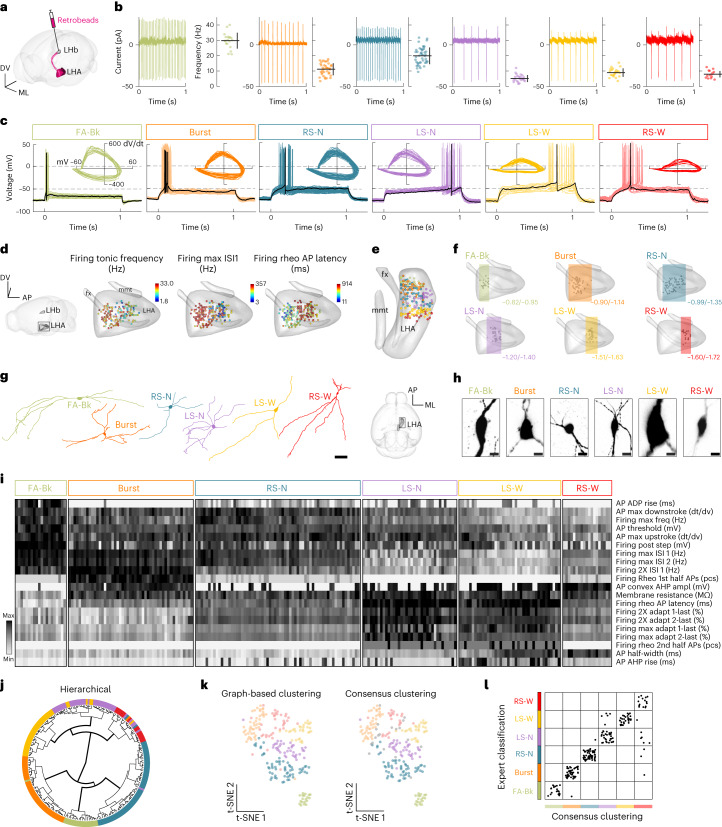
Fig. 1. Electrophysiological diversity of LHA–LHb neurons.
a, Strategy for retrograde labeling of LHA–LHb neurons. b, Cell-attached electrophysiological recordings reveal tonic firing of LHA–LHb neurons. Cell-attached traces from representative neurons (left) and the mean firing rate of the individual neurons (mean ± s.d.; right). nneuron = 230, from left to right nneuron = 20, 49, 65, 37, 40, 19 and nmice = 46 WT. c, Overlay of the individual whole-cell traces of firing at rheobase (Rheo) of all recorded LHA–LHb neurons. Black represents a trace from a representative neuron. Inset, phase-plane plots of the first AP at rheobase for all individual neurons. d, Electrophysiological properties reveal anatomical organization of LHA–LHb neurons. Dots represent recorded LHA–LHb neurons color coded by different electrophysiological parameters. e, Three-dimensional position of all recorded LHA–LHb neurons (color coded by cell type). f, Three-dimensional visualization of the A-P distribution of electrophysiologically characterized LHA–LHb neuron types. Bregma coordinates show the most anterior and posterior coordinates for each subtype. g, Reconstruction of representative dendritic morphologies of LHA–LHb neuron types. h, Images of representative soma morphologies of the LHA–LHb neuron types. i, Heatmap of 20 electrophysiological parameters selected based on PCA. Classification of LHA–LHb neuron types by expert classification, as in c. One column = one LHA–LHb neuron. j, Circular dendrogram for hierarchical clustering of LHA–LHb neurons. Color code, expert classification of LHA–LHb neurons. k, t-distributed stochastic neighbor embedding (t-SNE) plots of graph-based clustering (left), and consensus clustering (right). Color code, consensus clustering of LHA–LHb neurons. l, Agreement between the expert classification and the unsupervised consensus clustering of LHA–LHb neurons. All data acquired in male mice. Scale bar, 50 μm (g), 10 μm (h). See also Extended Data Figs. 1 and 2 and Supplementary Tables 1–3. nneuron = number of neurons; nmice = number of mice. WT, wild type; A-P, anteroposterior; M-L, medio-lateral; D-V, dorso-ventral; 2XRheo, two times the rheobase current injection; Max, maximal firing frequency; Mem, membrane intrinsic property; pcs, pieces.

