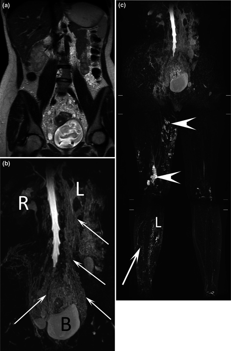
Fig. 7.
17-year-old female patient with chyluria and vaginal chylous discharge. Coronal T2-weighted MR image a demonstrated abnormal lymphatic vessels in the abdomen, pelvis, and retroperitoneum, which were better seen with (b) coronal MR lymphography with MIP reconstruction. Dilated lymphatic vessels (arrows) close to the bladder, ureters, colon, and uterine cervix were also demonstrated. R right kidney, L left kidney. This patient also presented with right lower limb lymphedema (L) and cystic lymphatic malformations (arrows) on coronal MR lymphography with MIP reconstruction (c)

