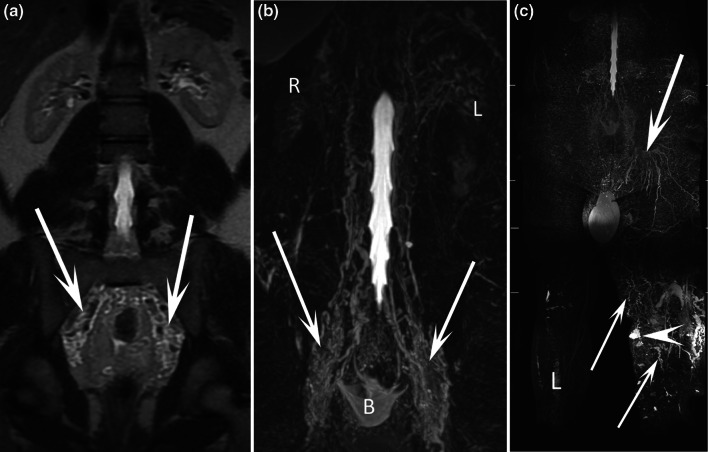
Fig. 8.
17-year-old male patient with chyluria, hydrocele, and bilateral lymphedema of the lower limbs. Coronal T2-weighted MR image (a) and coronal MR lymphography with MIP reconstruction b demonstrated widespread dilated lymphatic vessel development along the urinary tract from the retroperitoneum to the bladder (arrows). R right kidney, L left kidney. Coronal MR lymphography with MIP reconstruction (c) demonstrated major lymphatic dysplasia at the root of the left lower limb (arrows) and the presence of several cystic lymphatic malformations (arrowhead) of the soft tissues

