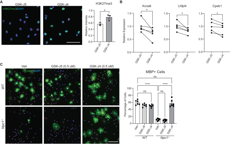
Fig. 6. Inhibiting H3K27 demethylase promotes oligodendrocyte differentiation in vitro.
Primary OPCs from WT and Npc1−/− mice were treated with the KDM6A and KDM6B inhibitor GSK-J4 (0.5 µM), the inactive control GSK-J5 (0.5 µM), or DMSO vehicle (Veh) for 4 days. A Npc1−/− cells treated with GSK-J5 or GSK-J4 and stained for H3K27me3 (green) and DAPI (blue). Intensity of H3K27me3 staining was measured relative to WT cells treated with GSK-J5. B Npc1−/− cells were treated with GSK-J5 or GSK-J4, and gene expression was quantified by qRT-PCR relative to cells treated with GSK-J5. Lines connect paired samples from biological replicate cultures. C Cells were stained for SOX10 (red) and MBP (green) to assess differentiation. Nuclei stained with DAPI (blue). The percentage of MBP-positive cells is quantified at the right. Scale bars = 50 µM (A) and 100 µM (C). Data are mean ± SEM (A, C). n.s. not significant, *p < 0.05, ****p < 0.0001 by two-tailed unpaired Student’s t-test (A), two-tailed paired Student’s t-test (B), and one-way ANOVA with Tukey post hoc test and multiple comparisons (C). A n = 3 biological replicates; t = 3.510; df = 4; p = 0.0247 (B) n = 5 biological replicates; t = 2.782, 3.479, 3.158; df = 4; p = 0.0497, 0.0254, 0.0343; (C) n = 6 biological replicates; F = 95.44; df = 5. Source data are provided as a Source Data file.

