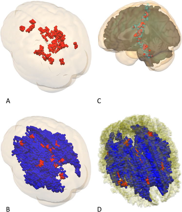
Figure 3.
This figure depicts voxels that were selected by comparing AIC statics of the 4 ageing models under study. Only voxels with sufficient model fit (Chi-square p > 0.05) exhibiting non-corrupt correlations between latent factors (r < 1 and r > − 1) were depicted within the confines of the skeletonized white matter (FA > 0.2). Voxels related to the cognitive mediation model are depicted in red while voxels of independent factor model are depicted in blue. (A) Depicts the caudal dorsal bird's-eye view of voxels related to the cognitive mediation model thresholded at a volume of 300 mm3. (B) Depicts the caudal dorsal bird's-eye view of voxels related to the cognitive mediation model and independent factor model thresholded at a volume of 300 mm3. (C) Depicts voxels related to the cognitive mediation model that were found within the confines of the postcentral /cerebellum white matter tract as described by the IIT Human Brain Atlas thresholded at a volume of 120 mm3. (D) Depicts the rostral dorsal bird's-eye view of both models within the confines of the cingular fibers as described by the IIT Human Brain Atlas thresholded at a volume 300 mm3.

