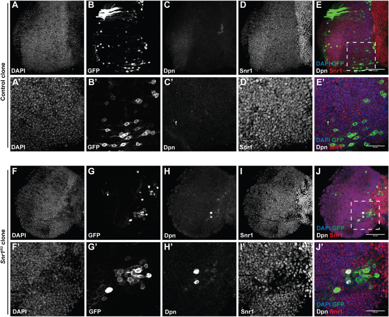
Fig. 4.
Cells expressing the neuroblast marker Deadpan are present in Snr1R3 clones in the adult optic lobe. Clones generated at 31 h after larval hatching (ALH). (A-E) Control MARCM clones marked with GFP. (A′-E′) Higher magnification images of region outlined in E. (F-J) Snr1R3 MARCM clone marked with GFP. (F′-J′) Higher magnification images of region outlined in J. In the merge, DAPI is in blue, GFP is in green, Dpn is in white and Snr1 is in red. Scale bars: 50 µm in E,J; 20 µm in E′,J′.

