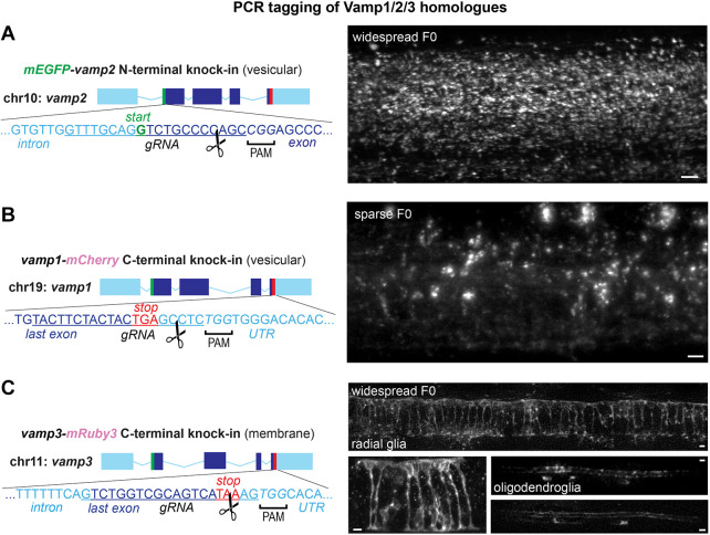
Fig. 3.
PCR tagging of related homologues Vamp1, Vamp2 and Vamp3. (A-C). Targeting additional vamp2 (N-terminal, A), vamp1 (C-terminal, B) and vamp3 (C-terminal, C) genomic loci. Representative widespread and sparse F0 larvae show vesicular mEGFP-Vamp2 and Vamp1-mCherry throughout the spinal cord and a membrane-like expression pattern of Vamp3-mRuby3 in radial and myelinating glial cells in the spinal cord. Scissors indicate predicted RNP cleavage site. Scale bars: 5 µm.

