. 2023 Apr 18;307(4):e222045. doi: 10.1148/radiol.222045

Figure 5:

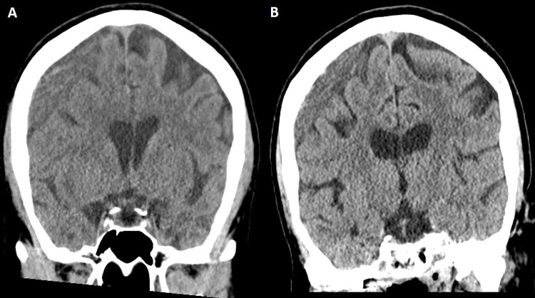
Case illustration of radiographic failure. (A) Head CT image in a 63-year-old woman shows a large right-sided chronic subdural hematoma (maximal thickness of 19 mm), which was discovered after the patient fell. Because of progressive headaches, the patient underwent a middle meningeal artery embolization procedure, which was uneventful. (B) Head CT image obtained at the 10-week follow-up (last available) shows minimal reduction of hematoma thickness, and the patient reported little improvement of headaches.

Case illustration of radiographic failure. (A) Head CT image in a 63-year-old woman shows a large right-sided chronic subdural hematoma (maximal thickness of 19 mm), which was discovered after the patient fell. Because of progressive headaches, the patient underwent a middle meningeal artery embolization procedure, which was uneventful. (B) Head CT image obtained at the 10-week follow-up (last available) shows minimal reduction of hematoma thickness, and the patient reported little improvement of headaches.