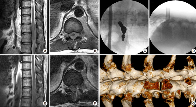
Fig. 3.
A patient with acute onset of both sides of paraplegia underwent full-endoscopic decompression surgery. (A, B) Preoperative magnetic resonance imaging (MRI) images reveal T10–12 epidural metastases and severe spinal cord compression. (C, D) Intraoperative fluoroscopic images show the optimal position for the interlaminar approach used in this case. (E, F) Postoperative MRI images demonstrate the tumor has been removed and the neural element was free. (G) Postoperative computed tomography scan showed the postlaminectomy site, where wide decompression was achieved.

