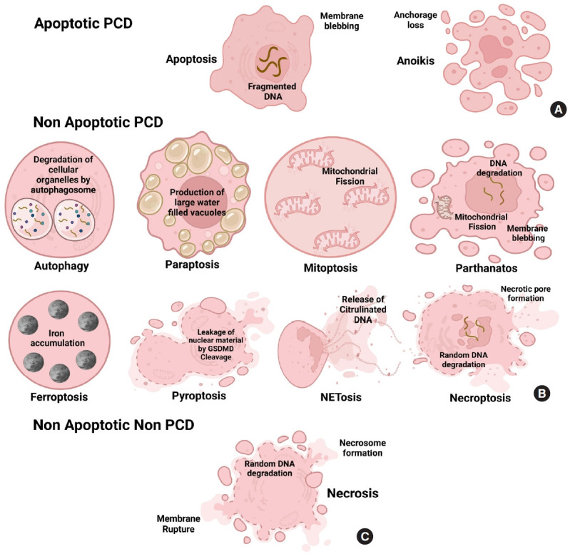
Fig. 2.

Morphology of different key cell death pathways. (A) Apoptosis and anoikis are classified under apoptotic programmed cell death (PCD). In apoptosis, nuclear DNA is fragmented, and the membrane is blebbed, while in anoikis, the cells lose their anchorage property to metastasize to other tissues. The morphological alteration in apoptotic non-PCDs is based on the presence of vacuoles, disruption of membrane integrity, organelle arrangement, chromatin density, and cell size. (B) It can involve mitochondria-mediated (mitoptosis, parthanatos), where mitochondrial integrity is disrupted, disabling the cell from producing energy for its survival; vacuole-mediated (autophagy, paraptosis), where the cell develops large hollow or fluid-filled vacuoles or cracks, iron-mediated (Ferroptosis), where cell accumulates iron inside to produce toxic effects, Immune factor-mediated (NETosis, pyroptosis) where immunological cascade controls the death of the cell or other various factor-mediated (necroptosis) that involves the change in oxidative stress, pH and formation of a necrotic cavity. (C) Nonprogrammed nonapoptotic form of cell death contains necrosis involving the spontaneous formation of necrosome and the release of cellular material that attack neighboring cells to go into necrosis. Triggered by toxins, physical injuries, and infections disrupting ionic pumps leading to Ca2+ influx, resulting in morphological alterations such as cytoplasmic swelling, consequential intracellular organelle loss with little to no chromatin condensation, and plasma membrane rupture.
