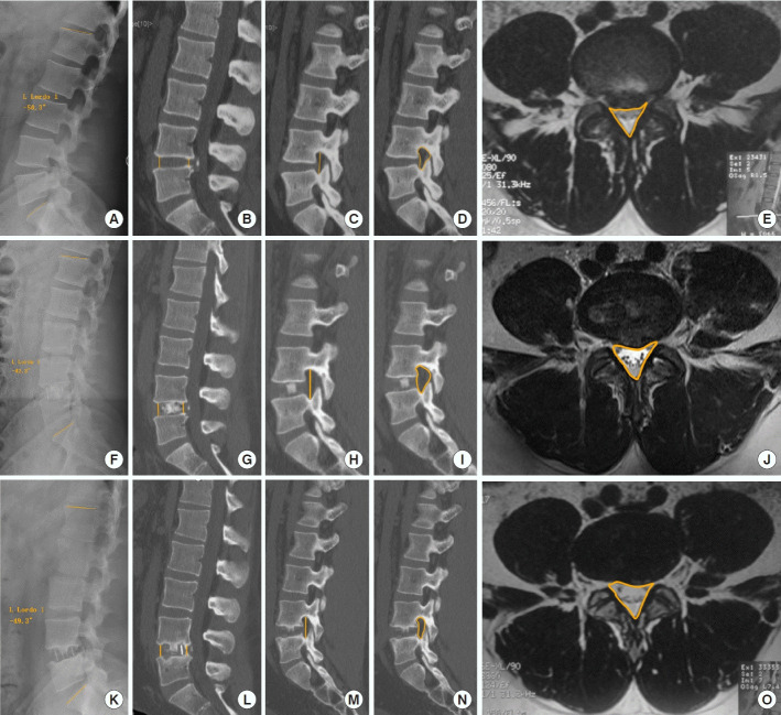
Fig. 2.
Radiographic evaluation. Measurement of cross-sectional area (CSA), anterior disk height (ADH), posterior disc height (PDH), foraminal height (FH), lumbar lordosis (LL), and the cross-sectional area of the intervertebral foramina (CSAF). Measurements of CSA, ADH, PDH, FH, and CSAF in the Picture Archiving Communication System (PACS) before the operation, at postoperative 1 day, and at last follow-up (A, F, and K). LL was measured in x-ray sagittal position, the head end measurement line was placed on the L1 superior end plate, the tail end measurement line was placed on the S1 superior end plate (B, G, and L). ADH and PDH were measured at the sagittal position in computed tomography (CT), the distance between the anterior/posterior edges of the upper vertebrae and the end plate of the lower vertebrae is called the anterior/posterior disk height (C, H, and M). FH was measured in the sagittal planes of the bilateral foramen in CT images. The length between the upper and lower edges is FH (D, I, and N). In the same plane of computed tomography CT, the foramen was outlined to read the CSAF (E, J, and O). CSA was measured in the axial sections of T2-weighted imaging. The central canal (including the thecal sac and epidural fat) was outlined in PACS to get the CSA.

