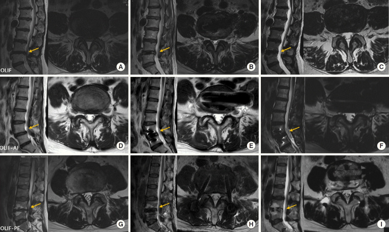
Fig. 5.
The magnetic resonance imaging (MRI) presentation in an operative patient. (A–C) The patient underwent OLIF. (D–F) The patient underwent OLIF-AF. (G–I) The patient underwent OLIF-PF. Three groups in MRI before the operation, at postoperative 1 day, and at last follow-up. The yellow arrow points to the sagittal segment corresponding to the axial MRI image.OLIF, oblique lateral interbody fusion; OLIF-AF, OLIF combined with anterolateral screw fixation; OLIF-PF, OLIF combined with percutaneous pedicle screw fixation.

