
FIG. 3.
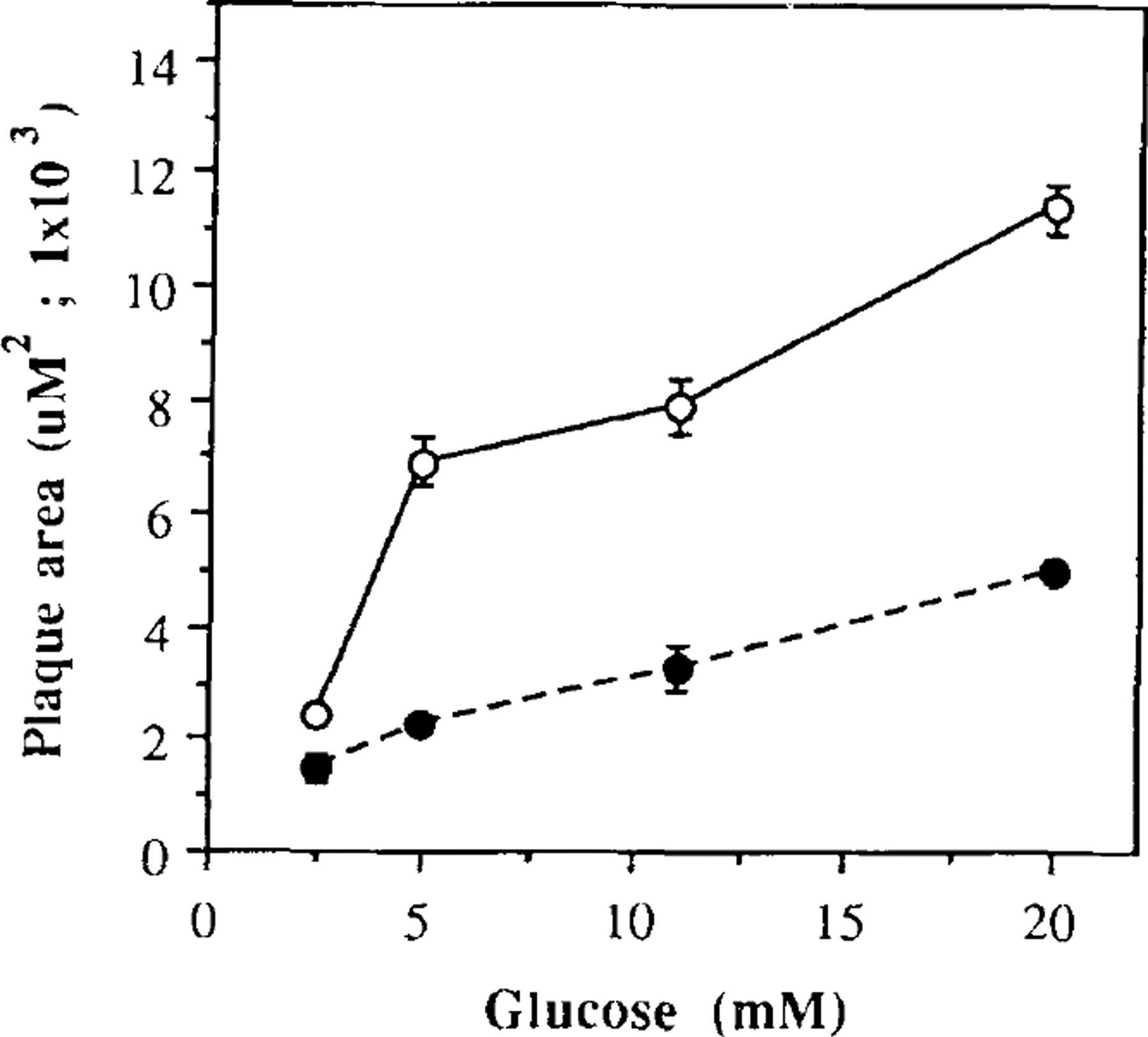
Mean plaque areas at various glucose concentrations. Dispersed islet cells from short-term (open circles) and long-term (filled circles) cultures were exposed to different glucose concentrations in the presence of insulin antiserum for 1 h. Datum points express the mean plaque area, which corresponds to the extension of the area of erythrocyte hemolysis. The values shown represent the mean ± SEM from three experiments. Each experiment was performed using pool of islets obtained from four rats.