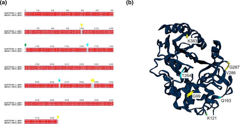
Fig. 3.
Nucleotide sequence of ADC-5 and its predicted structure. Alignment of putative ADC-5 cephalosporinase from AB341-IK15 and characterized ADC-5 AJ575184 (a). Structural predictions were done after amino acid alignment in MAFFT with ESPript. Characterized mutations are denoted with cyan arrows while novel mutations are highlighted with yellow arrows and K121R is highlighted with a green arrow as it is located in the binding pocket of ADC-5. Structural prediction of putative ADC-5AB341-IK15 using previously described ADC-5AJ575184, mutations in ADC-5AB341-IK15 are highlighted (b). Characterized mutations are highlighted in cyan while novel mutations are in yellow and green. G99A is located in the H2 α helices and K121R is a residue that makes up the binding pocket (highlighted in green).

