Fig. 3.
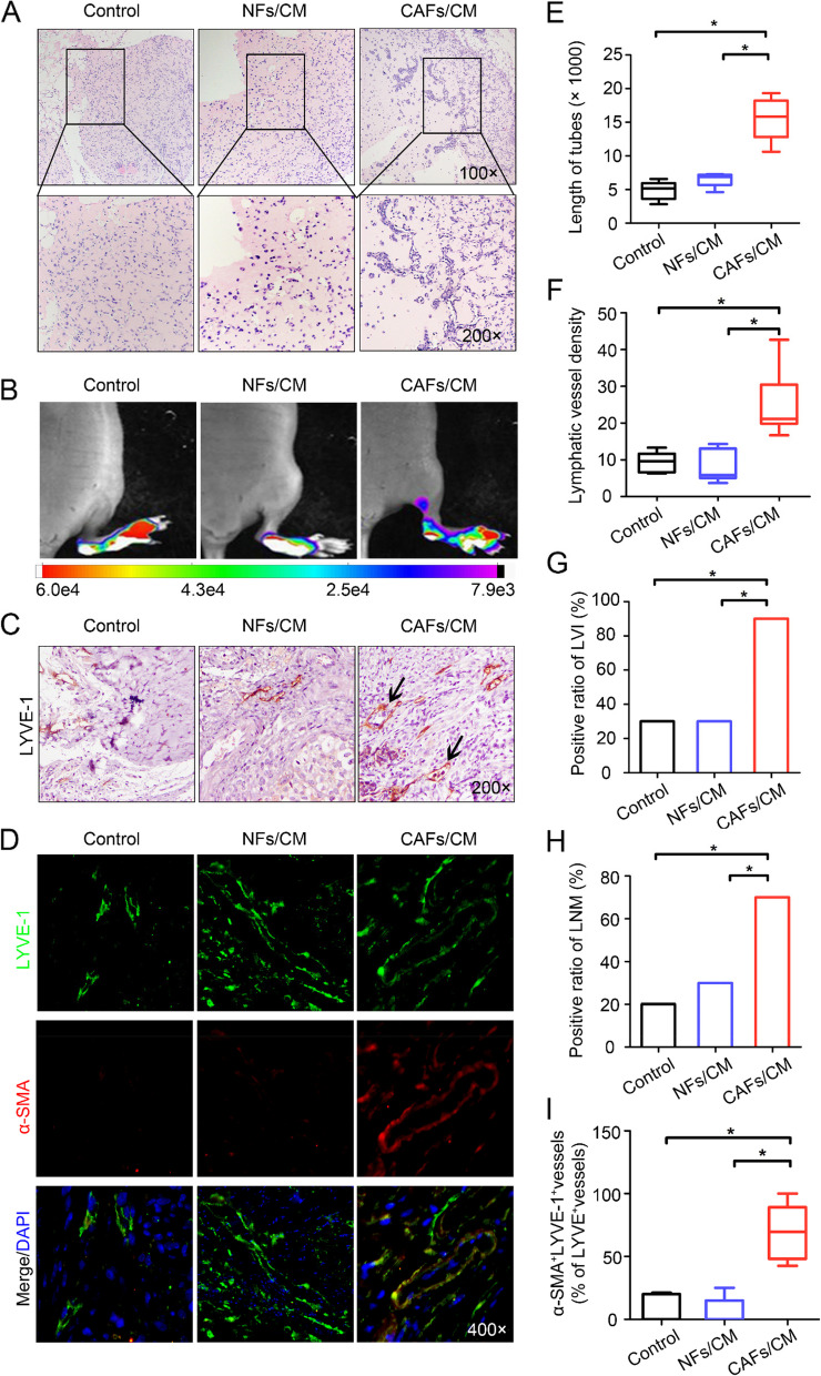
CAFs induce EndoMT and promote lymphangiogenesis in vivo. A Representative images showing tube formation in vivo (left panels: 100 × magnification; right panels: 400 × magnification). B In vivo fluorescence images of lymphatic metastasis (n = 10). C Staining of LYVE-1 in footpad tumours. Representative micrographs of positive staining are shown. Blank arrows indicate cancer cells that invaded the LVs. D Paraffin-embedded tumour sections from the experimental mice were stained with both anti-LYVE-1 (green) and anti-α-SMA (red) antibodies, and representative images are shown at 400 × magnification. E Statistical analysis showing the length of tube formation. The average tube length per field was calculated. F Statistical analysis of the LVD in footpad tumours. G Statistical analysis of lymphatic vessel invasion (LVI) in footpad tumours. H Ratio of metastasis-positive LNs to total dissected popliteal LNs in mice treated with the indicated CM. I Percentage of α-SMA+LYVE-1+ vessels among total LYVE-1.+ vessels in footpad tumours. *P < 0.05

