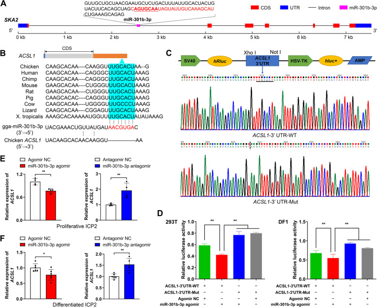
Fig. 7.
Validation of the ACSL1 gene as a direct target of gga-miR-301b-3p. A Schematic diagram of the genomic location of gga-miR-301b-3p. The sequences encoding the precursor gga-miR-301b-3p are shown, with the mature gga-miR-301b-3p sequences highlighted in red. B Complementary sequences of gga-miR-301b-3p and the 3′ UTR of the ACSL1 gene of different species. The seed region of gga-miR-301b-3p is highlighted in red; potential gga-miR-301b-3p-binding sites in the 3′ UTR of the ACSL1 gene are shown in blue. C Construction of dual-luciferase reporter vectors for the validation of gga-miR-301b-3p targeting the ACSL1 gene. WT: wild-type vector; mut: mutant vector; hRluc: Renilla luciferase; hluc + : firefly luciferase. D Validation of the interaction between gga-miR-301b-3p and the 3′ UTR of the ACSL1 gene by a dual-luciferase reporter assay in 293 T cells and DF1 cells. E Relative ACSL1 mRNA expression in gga-miR-301b-3p-overexpressing and -knockdown proliferative chicken abdominal preadipocytes. The relative gene expression levels are shown as fold changes compared with that in the agomir NC and antagomir NC groups, respectively. The same below. F Relative ACSL1 mRNA expression in gga-miR-301b-3p-overexpressing and -knockdown differentiated chicken abdominal preadipocytes. *P < 0.05, **P < 0.01

