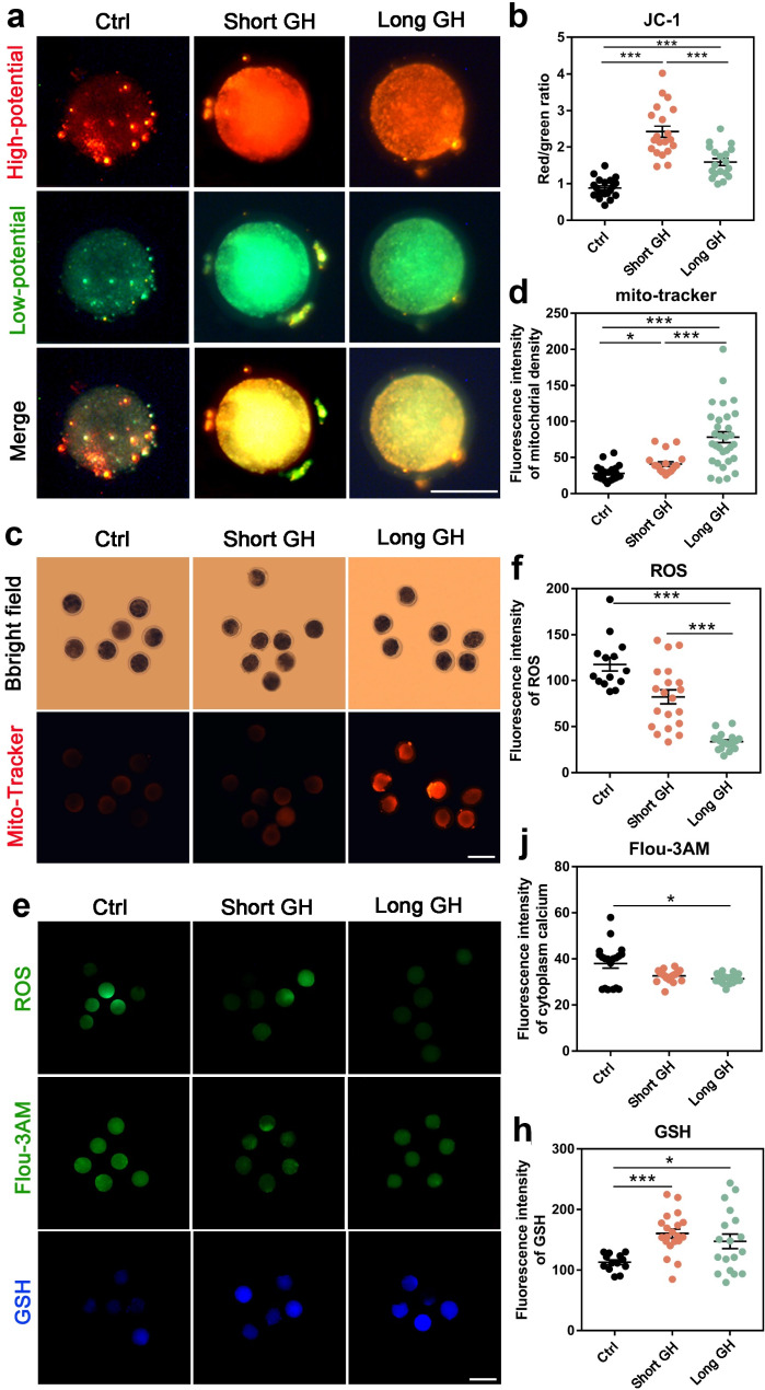
Fig. 3.
Detection of the relative indexes of lamb oocyte quality in trial 3. a The representative images of staining for MMP in lamb oocytes (scale bar = 50 μm), Red indicates high Δφm, while green indicates low Δφm. b The quantification of MMP levels detected by fluorescence intensities. c The representative images of mitochondrial masses staining (Mito-Tracker) in lamb oocytes (scale bar = 100 μm). d The quantification of mitochondrial masses detected by fluorescence intensities. e The representative images of staining for ROS, cytosolic Ca2+ (Floµ-3AM) and GSH content in lamb oocytes from top to bottom (scale bar = 100 μm). f-j The quantification of levels of ROS, cytosolic Ca2+ and GSH content detected by fluorescence intensities. Ctrl, control; long GH, long GH treatment (for 5 days); short GH, short GH treatment (for 2 days). *P<0.05; ***P<0.001

