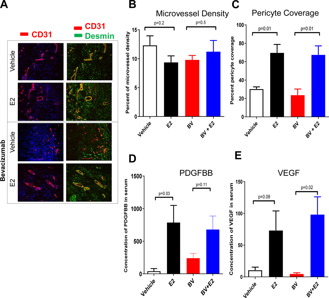
Figure 3. Estrogen increases pericyte vessel coverage and pro-angiogenic growth factor secretion in NSCLC xenografts.
Representative of immunofluorescence staining (20X) of CD31 (red), desmin (green) and nuclei (blue) using immunofluorescence microscopy. A minimum of 4–5 microphotograph (20X) for each sample (n=4/group) were collected (A-B). Quantification of microvessel density (C) and pericyte coverage (D) at tumor progression (100mm3) was performed. Vessels were considered pericyte covered if more than 50% of CD31+ vessel co-localized with desmin. Data are graphed as mean ± SEM. Increased circulating angiogenic factors, PDGFBB and VEGF were measured during tumor growth in presence of estrogen. Mouse PDGFBB (E) and VEGF (F) serum levels were quantified using the Bio-rad multiplex bead assay. For each sample (n=4/group), blood from tumor bearing mouse was drawn by cardiac puncture. The results were plotted as average of the duplicate and graphed as mean ± SEM. Mice were treated with bevacizumab (BV) 10mg/kg in presence or absence of estrogen.

