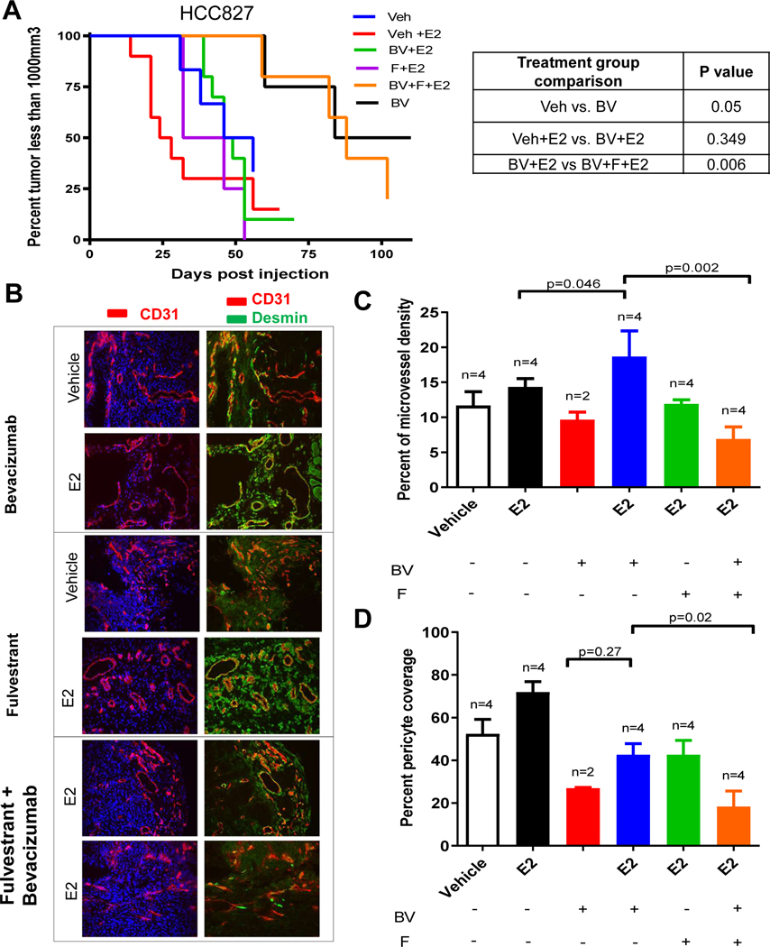
Figure 5. Estrogen receptor blockade sensitizes NSCLC tumors to anti-VEGF therapy.
Nude ovariectomized mice were primed with slow releasing estrogen (80pg/ml) tube. HCC827 xenograft mice were treated with bevacizumab (BV) 10mg/kg twice in a week, and fulvestrant (F) 200mg/kg, once in a week in presence of estrogen. Kaplan-Meier plot showing survival distribution of HCC827 xenograft treated with BV, F, or estrogen (A). The log-rank (Mantel-Cox) test was used to compare the statistical differences among the groups. Representative images of immunofluoroscence staining (200X) of CD31 (red), desmin (green) and nuclei (blue) using immunofluorescence microscopy were collected (B). A minimum of 4–5 microphotograph (200X) for each sample (n=4/group) were collected. BV alone group had two tumor bearing mice. Quantification of microvessel density (C), pericyte coverage (D), and CD11b+ myeloid cells (E) by staining was measured.

