FIGURE 1.

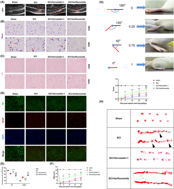
Dihydroorotate dehydrogenase can improve tissue damage of neurons after spinal cord injury and promote functional recovery. (A) Representative MR images of the spinal cord of rats in each group at 4 weeks after spinal cord injury, with injury areas marked by red boxes. (B) Nissl staining and Nissl staining for partial enlarged images. The number of motor neurons in the anterior horn of spinal cord was observed and analyzed by Nissl staining. The arrow represents neurons containing Nissl bodies (n = 10 for each group, scale bar = 20 μm). (C) HE staining images of representative spinal cord cross‐sections of each group (n = 10 for each group, scale bar = 50 μm). (D, E) NF (Green) and GFAP (Red) immunofluorescence staining. Each group reflects the functional status of nerve fibers, the ability to maintain continuous nerve fibers and inhibit astrocyte proliferation. (Scale bar: 100 μm. Image J software is used to measure the density of GFAP and NF in three randomly selected fields of vision.) (F) BBB scores were used to assess changes in hind limb motor recovery at 3, 7, 14, 21, and 28 days after spinal cord injury (n = 10 for each group). (G) Schematic score representation of the evaluation of ankle flexibility and the ankle flexibility score of each group (n = 10 for each group). (H) Representative images of footprints from each group. The hindlimbs were displayed in red (n = 10 for each group). (All the data are expressed as means ± SD, one or two‐way ANOVA followed by Tukey's post hoc test was applied; *p < 0.05, **p < 0.01, ***p < 0.001 vs. SCI; # p < 0.05, ## p < 0.01, ### p < 0.001 vs. SCI + ferrostatin‐1; ^ p < 0.05, ^^ p < 0.01, ^^^ p < 0.001 vs. SCI + teriflunomide.)
