
FIGURE 6.
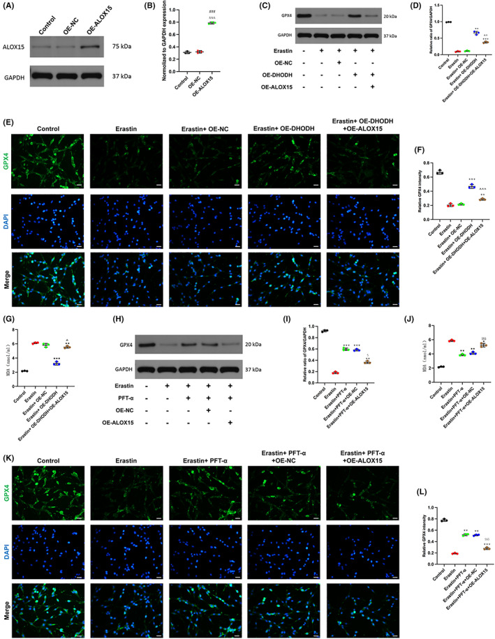
Dihydroorotate dehydrogenase inhibits neuronal ferroptosis after spinal cord injury by inhibiting P53/ALox15. (A, B) The expression of OE‐ALOX15 in transfected pc12 cells. (C, D) Western blot detection and quantification of GPX4 expression under treatment with erastin or up‐regulation of DHODH or up‐regulation of ALOX15. (E, F) The expression of GPX4 was detected by immunofluorescence staining and quantitative statistical analysis (scale bar = 50 μm, the relative content of GPX4 was calculated by Image J software). (G) MDA content in each group was detected by MDA kit. (H, I) After treatment with P53 inhibitor, the expression of GPX4 was detected by Western blot and quantitatively counted. (J) Detecting the MDA content of each group after treatment with P53 inhibitor. (K, L) Immunofluorescence staining showed that inhibition of P53 activity significantly promoted the expression of GPX4, and overexpression of ALOX15 reversed this effect (scale bar = 50 μm, the relative content of GPX4 was calculated by Image J software). (All the data are expressed as means ± SD, n = 3, one‐way ANOVA followed by Tukey's post hoc test was applied *p < 0.05, **p < 0.01, ***p < 0.001 vs. Erastin; # p < 0.05, ## p < 0.01, ### p < 0.001 vs. Control; ^ p < 0.05, ^^ p < 0.01, ^^^ p < 0.001 vs. Erastin + OE‐DHODH; & p < 0.05, && p < 0.01, &&& p < 0.001 vs. OE‐NC; $ p < 0.05, $$ p < 0.01, $$$ p < 0.001 vs. Erastin + PFT‐α.)