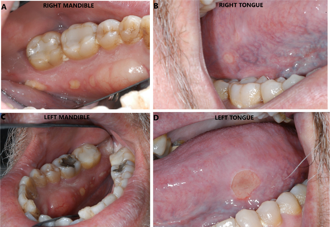
Figure 1.
A: A 4×5 mm area of exposed bone with trailing smaller lesions seen on the right postero-lingual mandibular alveolus.
B: A 2×2 mm ulceration seen on the right postero-lateral border of the tongue, secondary to trauma from the sharp exposed bone.
C: A 3×3 mm area of exposed bone with trailing smaller lesions seen on the right postero-lingual mandibular alveolus.
D: A 8×6 mm ulceration seen on the left postero-lateral border of the tongue, secondary to trauma from the sharp exposed bone.

