Figure 3.
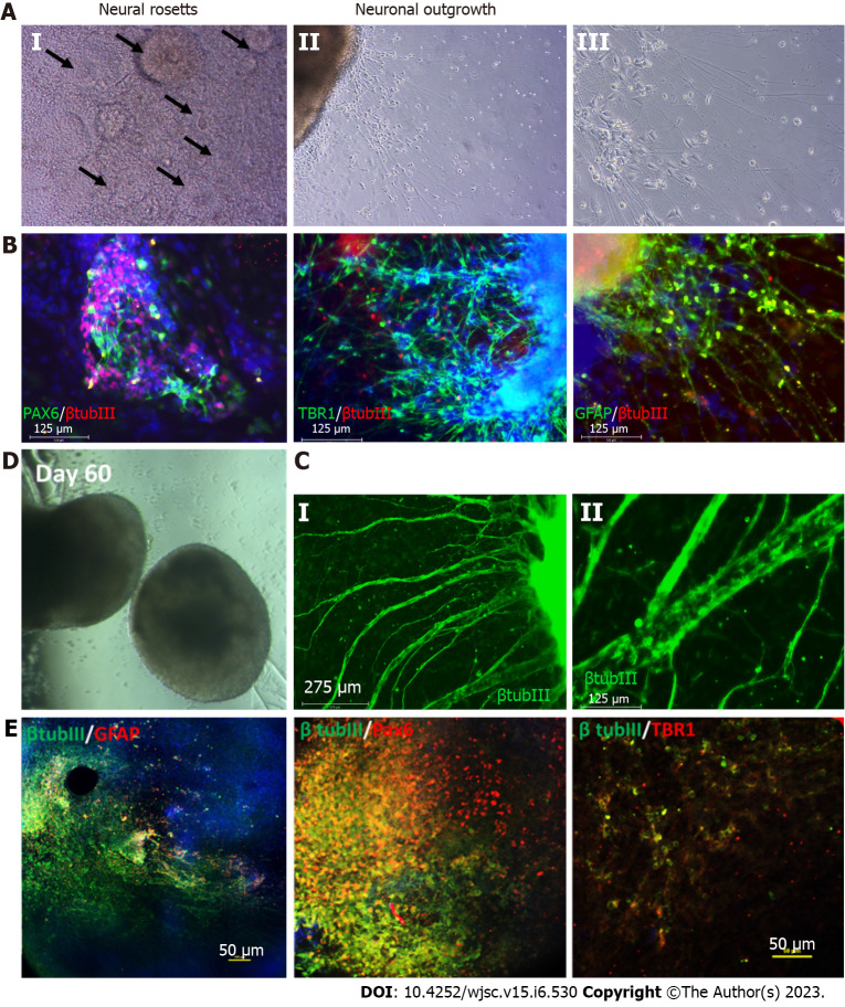
Characterization of cortical organoids for neural and astrocyte marker expression. A: Brightfield images showing the neural rosettes and neuronal outgrowth from the organoids replated to an attachment plate at day 35 of differentiation; B: Resulting immunocytochemistry analysis of neural marker paired box 6 (PAX6), cortical deep layer VI marker T-box brain transcription factor 1 (TBR1), astrocyte marker glial fibrillary acidic protein (GFAP) co-stained with common neural marker β tubulin III, scale bar 125 µm; C: Immunostaining at later stage of the replating showing thick axon like extensions from the organoids, scale bar: 275 µm; D: Brightfield images of the day 60 cortical organoids; E: Confocal images of the day 60 organoids showing astrocyte marker GFAP, neural marker PAX6, cortical deep layer VI marker TBR1 co-stained with common neural marker β tubulin III, scale bar: 50 µm.

